Buscar
- Nuestro Centro
- Entorno asistencial Teknon
- Institutos de tratamiento especializado
- Campus TeknonEl área de Consultorios dispone de 2 edificios de consultas con 150 despachos médicos, con conexión directa con la clínica y acceso directo al parking.
- Especialidades
- Unidades especializadas
- Medicina y Cirugía sin sangre
- Reproducción Asistida
- Unidad de Atención al Lesionado de Tráfico
- Unidad de Endoscopia y Pruebas Funcionales Digestivas
- Unidad de Enfermedades Inflamatorias y Autoinmunes
- Unidad de Epilepsia y Cirugía de Epilepsia
- Unidad de Medicina Marítima
- Unidad de Neumología
- Unidad de Obstrucción Respiratoria Nasal
- Unidad de Pediatría y Cirugía pediátrica
- Unidad de Síndromes de Sensibilización Central
- Unidad de Tratamiento del Dolor
- Unidad de VPH y Displasia anal
- Unidad del Aliento
- Unidad del Parkinson y Trastornos del movimiento
- Unidad del Sueño
- Especialidades
- Cardiología y cirugía cardiovascular
- Cirugía general
- Cirugía estética
- Cirugía maxilofacial
- Clínica del Dolor
- Gastroenterología
- Ginecología y obstetricia
- Medicina Interna
- Neumología
- Neurocirugía
- Oncología
- Otorrinolaringología
- Pediatría y cirugía pediátrica
- Traumatología
- Urología
- Todas las especialidades
- Localice un especialistaEn nuestro campus ejercen más de 400 especialistas de primera línea, con formación internacional en cada una de sus áreas y atención al paciente en varios idiomas.
- Unidades especializadas
- Área diagnóstica
 Diagnóstico cardiaco
 Pruebas diagnósticas
 Chequeos médicos This link will open in a pop-up window.
- General This link will open in a pop-up window.
- Completo This link will open in a pop-up window.
- Completo Plus This link will open in a pop-up window.
- Viajeros This link will open in a pop-up window.
- Deportivo This link will open in a pop-up window.
- Cardiológico This link will open in a pop-up window.
- Empresa This link will open in a pop-up window.
- Exclusividad/Club Teknon
- La mejor atención
- La mejor asistencia
- Otros servicios
- Actualidad
- NoticiasConoce qué está pasando en Centro Médico Teknon. Consulta nuestra sección de noticias. Te interesará.
- Contenidos de saludTu salud es lo que cuenta
- AgendaPuedes encontrar todos los eventos que hemos organizado sobre salud y aquellos temas de actualidad que te pueden interesar. Accede a nuestra agenda de actividades.
- El Blog de Centro Médico TeknonLa salud, la ciencia y la medicina responden a la racionalidad, el empirismo, los resultados y las cifras. Pero, ¿qué hay de las personas? El factor humano es una parte esencial en la medicina que, algunas veces, queda en segundo plano. This link will open in a pop-up window.
Garces Gatnau Joan Ramón
Melanoma. The most malignant skin cancer
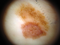
Melanoma is the type of skin cancer that has increased the most in recent years and is potentially the most aggressive. It originates with the cancerous transformation of melanocytes, which are cells found in normal skin that provide each person with their own skin colour. Melanoma can appear on a pigmented lesion (melanocytic nevi) or on healthy skin. Any pigmented lesion that changes in colour and/or in appearance should be checked by a dermatologist.
It is considered high risk cancer because melanoma is a tumour that can spread and produce metastasis very quickly. If melanoma is diagnosed early, the treatment simply involves surgery. That is why an early diagnosis with digital dermatoscopic monitoring is so important as it reduces the possibility of death.
What does skin cancer screening involve?
The Dermatekpigmented lesion unit offers the following services:
- An annual pigmented lesion check-up with dermatoscopy
- Body mole mapping with digital dermatoscopy: FotoFinder dermoscope®
- Follow-up, diagnosis and treatment of patients with melanoma
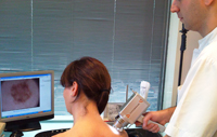
Digital Dermatoscopy
Digital dermatoscopy or epiluminescence microscopy with FotoFinder dermoscope® is currently the standard of excellence in monitoring pigmented lesions and in the early diagnosis of melanoma and all other malignant skin tumours.
Digital dermatoscopy improves the accuracy of a skin cancer diagnosis and prevents unnecessary extirpation and biopsies of benign lesions. Furthermore, it can be used to perform periodic side-by-side comparisons of pigmented lesions to detect even the slightest change that may suggest malignization. This reduces the chances of death. For more information, you can view the Atlas of Practical Dermatoscopy.
| Morning | Afternoon | |
|---|---|---|
| Monday | 9.00 - 15.00 h | 15.00 - 20.00 h |
| Tuesday | 9.00 - 15.00 h | 15.00 - 20.00 h |
| Wednesday | 9.00 - 15.00 h | 15.00 - 20.00 h |
| Thursday | 9.00 - 15.00 h | 15.00 - 20.00 h |
| Friday | 9.00 - 15.00 h | 15.00 - 20.00 h |
- Nuestro Centro
- Entorno asistencial Teknon
- Institutos de tratamiento especializado
- Campus Teknon
- Especialidades
- Unidades especializadas
- Especialidades
- Localice un especialista
- Área diagnóstica
- Exclusividad/Club Teknon
- La mejor atención
- La mejor asistencia
- Otros servicios
- Actualidad

Carrer de Vilana, 12, 08022 Barcelona
932 906 200 / 900 301 013