Buscar
- Nuestro Centro
- Entorno asistencial Teknon
- Institutos de tratamiento especializado
- Campus TeknonEl área de Consultorios dispone de 2 edificios de consultas con 150 despachos médicos, con conexión directa con la clínica y acceso directo al parking.
- Especialidades
- Unidades especializadas
- Medicina y Cirugía sin sangre
- Reproducción Asistida
- Unidad de Atención al Lesionado de Tráfico
- Unidad de Enfermedades Inflamatorias y Autoinmunes
- Unidad de Epilepsia y Cirugía de Epilepsia
- Unidad de Medicina Marítima
- Unidad de Neumología
- Unidad de Obesidad
- Unidad de Obstrucción Respiratoria Nasal
- Unidad de Pediatría y Cirugía pediátrica
- Unidad de Síndromes de Sensibilización Central
- Unidad de Tratamiento del Dolor
- Unidad del Virus del Papiloma Humano
- Unidad del Aliento
- Unidad del Parkinson y Trastornos del movimiento
- Unidad del Sueño
- Especialidades
- Cardiología y Cirugía Vascular
- Cirugía general
- Cirugía estética
- Cirugía maxilofacial
- Clínica del Dolor
- Gastroenterología
- Ginecología y obstetricia
- Medicina Interna
- Neumología
- Neurología y Neurocirugía
- Oncología
- Otorrinolaringología
- Pediatría y cirugía pediátrica
- Traumatología
- Urología
- Todas las especialidades
- Localice un especialistaEn nuestro campus ejercen más de 400 especialistas de primera línea, con formación internacional en cada una de sus áreas y atención al paciente en varios idiomas.
- Unidades especializadas
- Área diagnóstica
 Diagnóstico cardiaco
 Pruebas diagnósticas
 Chequeos médicos This link will open in a pop-up window.
- General This link will open in a pop-up window.
- Completo This link will open in a pop-up window.
- Completo Plus This link will open in a pop-up window.
- Viajeros This link will open in a pop-up window.
- Deportivo This link will open in a pop-up window.
- Cardiológico This link will open in a pop-up window.
- Empresa This link will open in a pop-up window.
- Exclusividad/Club Teknon
- La mejor atención
- La mejor asistencia
- Otros servicios
- Actualidad
- Comunicación
- AgendaPuedes encontrar todos los eventos que hemos organizado sobre salud y aquellos temas de actualidad que te pueden interesar. Accede a nuestra agenda de actividades.
- El Blog de Centro Médico TeknonLa salud, la ciencia y la medicina responden a la racionalidad, el empirismo, los resultados y las cifras. Pero, ¿qué hay de las personas? El factor humano es una parte esencial en la medicina que, algunas veces, queda en segundo plano. This link will open in a pop-up window.
Bardají Bofill Manel
- Nuestros servicios
Embryology
The development of the pharynx in the embryo determines the appearance of the main endocrine glands of the head and neck, arising from evaginations that we call pouches. These coincide on the outside with the system of pharyngeal arches, whose skeletal, nervous and arterial derivatives are shown in Table 2. These arches separated by grooves give the embryo the appearance of the gills of the fish, but at no time do they coincide outside with the interior as it happens in them.
The 1st and 2nd pharyngeal bags will give rise to the tympanic tube recess, from which the middle ear and the eustachian tube will be formed, the ventral component of the 2nd will give rise to the intramygdalin fossa. The dorsal wing of the 3rd pouch will give rise to the lower parathyroid, the ventral wing from the 3rd pouch to the thymus. The 4th pouch originated, its dorsal component will form the upper parathyroid, its ventral wing is actually the 6th bag or last gill body that supposes constitutes the interfollicular cells of the thyroid. This bilateral system leaves in its middle zone the odd tubercle and the hypobranchial eminence, which originate the tongue and thyroid.
Once these structures are outlined, to understand the formation of branchial cysts, we need to know a structure called the cervical sinus. The growth of the arches is different, so that the 1st and 2nd develop a lot, leaving the 3rd and 4th arches submerged in a shallow ectodermal fossa that we call cervical sinus. Likewise, the epipericardial crest rises enclosing the cervical sinus more deeply, from this crest the mastoid sternocleid muscle, the trapezius and the infrahoid muscles and the floor of the mouth will be formed, likewise the spinal and hypoglossal nerves, that is, these structures are not cranial, but were incorporated into the skull in the evolution of mammals.
The approximate area of the cervical sinus with triangular shape, is determined by derivatives of the epipericardial crest, the sternocleidomastoid, the trapezius, the infrahoid musculature and the floor of the mouth, the elements derived from the hyodes, thyroid and cricoid cartilage arches are within the cervical sinus, the hypoglossal nerves and their descending loop are always posterior to the cervical sinus.
In fact the growth of the 2nd arch is so important that it protrudes covering the gill slits as it happens in ossified fish, also determines that in muscular derivative of this arch, the platysma covers these structures, over time the cervical sinus is detached from the surface by closing the external orifice, the sinus is obliterated and normally leaves no remains.
 sinus in the embryonic period and its position in the adult a) cervical sinus b) cervical sinus in the adult c) epipericardial crest.
- Fistulas, sinuses and branchial cysts
They are congenital lesions due to the persistence of the cervical sinus, being formed by ectodermal epithelial tissue, if connections are maintained with the surface we will find a fistula that drains intermittently to the outside somewhere on the anterior surface of the mastoid sternocleid. If the connection is with the interior, with the bag system it allows us to classify them into connections with the second, third, fourth or sixth.
The connection with the 2nd pouch determines that from the cyst the fistula ascends following the carotid sheath, in front of the internal carotid and over the bifurcation( 3 arch) until it reaches the hypoglossal nerve turns on it because it is a derivative of the epipericardial crest and horad the pharyngeal wall leading to the fossa to intratonsillar (2nd arch).
The connection with the 3rd pouch passes behind the internal carotid veers over the hypoglossum and descends towards the pharynx entering it in the region of the piriform sinus at the level of the thyrohoid membrane more cranial than the superior laryngeal branch (4th arch). The connections with bag 4 and 6 are theoretically possible but would imply that after turning on the hypoglossal they descend to the root of the cuelo or to the anterior mediastinum.
Gill fistulas are always present at birth. They can be unilateral or bilateral. The external opening sits in the middle or lower third of the mastoid sternocleid muscle. Branchial cysts usually manifest in adults, in very few cases in children. They are usually 4 - 5 times more frequent than gill fistulas. They are located on the superficial face of the carotid sheath, deep with respect to the anterior edge of the sternocleidomastoid, at the height of its middle third. They appear as smooth masses more or less rounded, fluctuating, soft and painless and with some lateral mobility. It is necessary to establish the differential diagnosis with lymphadenopathy, hygroma, lipomas and hemamgiomas.
Both fistulas and gill cysts can become infected. When this happens, it should be treated with antibiotics before making its definitive treatment, being necessary in some cases to proceed to its evacuation.
We must complete the examination with a fine needle aspiration puncture, which will inform us of cuboid epithelial cells characteristic of the cyst wall, and computerized axial tomography that will define their relationships and the presence of accompanying lymphadenopathy.
The treatment is always surgical, for branchial cysts, a transverse incision is made following a fold of the skin of the neck. This excision is not usually easy; On the contrary, it requires all the care required by neck surgery at the carotid crossroads.
For the removal of fistulas, the Sistrunk stepped incision method is used. Two spindle incisions are drawn around the external fistulous opening and the fistula is dissected as high as possible. Another incision is drawn higher up to complete its detachment and complete excision. Sometimes 3-4 stepped incisions are necessary.
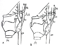 Relationships of the cysts of 2nd (A) and 3rd (B) arch with vascular, nervous and pharyngeal structures.
Common carotid PC, internal carotid IC, external carotid EC, LS superior laryngeal nerve,12 hypoglossal nerve,9
glossopharyngeal nerve.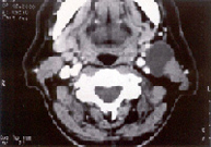 Quiste branquial de 2º arco, cubierto por
el músculo platisma, en lateral el ECM
y en profundidad la yugular interna y la carótida.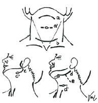 Surgery a) Central Superior, access to the Thyroglossal Cyst a') If Fistula is added to the Thyroglossal Cyst, it requires staggered incisions b) Central Inferior in Tie, incision to access the Thyroid and Parathyroid c) Parotectomy incision, suitable for cyst of 1st arch c') If Fistula of the first arch is added d) Optimal high lateral to access carotid triangle, Brachial cyst e) This prolongation allows adequate control for larger laterocervical lesions d ́) Fistula 2nd arch stepped incision
 - Congenital atrial fistulas
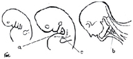
Abnormalities of the first cleft, also called congenital atrial fistulas make up at least 10% of the total. The average age of presentation is around 3 years. The malformation is located in an area comprising the external auditory canal, parotid, angle of the jaw, retroauricular region, submaxillary and upper cervical region, are due to the persistence or lack of obliteration of the first branchial arch or an incomplete fusion of the different auricular tubercles. It is not believed that these anomalies are related to the much more frequent pretragal cysts, which can reach the periosteum of the temporal bone, without any connection to the external auditory canal. The place where they manifest most frequently is the upper laterocervical region, followed by the parotid and auricular.
According to their location and embryological origin, they are classified into three groups:
- Preauricular fistulas between the mouth corner and the swallow.
- Preauricular fistulas located in front of the ascending root of the hellix and directed to the external auditory canal.
- Sacciform depressions or small blind fistulas that can be located anywhere in the pinna
Another classification is that of Work, which relates them to the external auditory canal and the facial nerve. Seria type I (Horizontal) of alteration is the simplest and consists of duplicity of the duct, appearing as fistula or preauricular ectodermal cyst are usually on the facial nerve. Type II (Vertical) presents in the form of an irregular mass of about 2-3 cm, with size fluctuations, frequent mucous drainage and occasional infection, extending from the angle of the jaw, sub- or retroauricular region to the internal auditory canal through the parotid, either medial or lateral to the facial nerve. Its treatment would require parotidectomy incision to identify the facial nerve and avoid its injury.
- Thyreoglossus duct cyst
It is a frequent malformation and constitutes 40% of cervical congenital anomalies They are due to the persistence of the tyreoglossal duct, which communicates the pharyngeal pouch above the foramen caecum of the tongue with the thyroid isthmus.
The descent of the thyroid gland to the trachea lengthens the tyreoglossal duct, which ends up obliteration, the failure of this process is manifested by the formation of cysts with or without external fistula. Clinically they are a cystic tumor, in the midline of the neck at the level of the small thyrohoid menbrana that ranges from that of a walnut to the egg of a pigeon, smooth surface, soft consistency, not adhered to the skin, but yes to the hyoid bone, painless on palpation and that moves with swallowing movements and when removing the tongue. They are asymptomatic, except when infected, an eventuality that usually occurs in more than 50% of cases. The infection usually occurs from the oropharyngeal cavity, which manifests itself at the level of the cyst by rapid increase in size, pain, imprecise delimitation on palpation and alteration of the normal characteristics of the skin that covers it. The only subjective symptom is usually pain when swallowing, but, if it is intralingual, it can produce dysarthria and dysphagia. Through conservative medical treatment, the situation returns to normal, although most often it fistuples. The external orifice of the fistula can be located along the entire anterior midline, that is, from the hyodes bone to the suprasternal hollow. The differential diagnosis must be established with midline dermoid cyst, lipomas and ectopic thyroid.
We must complete the exploration with a fine needle aspiration puncture, which will inform us of cuboid epithelial cells characteristic of the cyst wall, and the computerized axial tomography that will define its relationships with the hyodes.
The treatment is always surgical By means of a transverse incision and if it is a fistula two incisions in spindle that cover the fistulous orifice, the cyst or fistula is detached to the hyodes bone and as the central portion of the bone is always involved in this process, its middle portion is resected and the rest of the fistula is released to the base of the tongue. To facilitate this last maneuver, the anaesthetist inserts his finger into the pharynx and bulges its wall outwards. When the fistula is located distally, the procedure of stepped incisions of Sistrunk is followed.
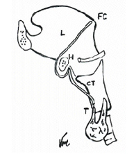 Path of the Thyreoglossus Duct : CT Tireoglossus
duct starts at FC Foramen Cecum, follows the L Tongue,
turns over the Hyoid H and ends in Thyroid T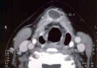 Cervical CT image, thyroglossal cyst, its central disposition and
its relationships with the hyoid bone can be seen
| Morning | Afternoon | |
|---|---|---|
| Monday | 09.00 - 13.00 h | 16.00 - 20.00 h |
| Tuesday | 09.00 - 13.00 h | 16.00 - 20.00 h |
| Wednesday | 09.00 - 13.00 h | 16.00 - 20.00 h |
| Thursday | 09.00 - 13.00 h | 16.00 - 20.00 h |
| Friday | 09.00 - 13.00 h | 16.00 - 20.00 h |
- Nuestro Centro
- Entorno asistencial Teknon
- Institutos de tratamiento especializado
- Campus Teknon
- Especialidades
- Unidades especializadas
- Especialidades
- Localice un especialista
- Área diagnóstica
- Exclusividad/Club Teknon
- La mejor atención
- La mejor asistencia
- Otros servicios
- Actualidad
- Comunicación
- Agenda
- El Blog de Centro Médico Teknon

Carrer de Vilana, 12, 08022 Barcelona
932 906 200