


- Cuadro médico
- Especialidades
- Unidades especializadas
 Instituto del Corazón
 Unidad de Obesidad
 Instituto Oncológico
 Unidad de Medicina y Cirugía sin sangre
 Instituto de Neurociencias
 Unidad de Atención al Lesionado de Tráfico
 Instituto de Neumología
 Unidad de Medicina Marítima
 Instituto de Terapia Regenerativa Tisular
 Unidad de Tratamiento del Dolor
 Clínica del Tenis
 Unidad de Enfermedades Inflamatorias y Autoinmunes
 Unidad de Reproducción
Asistida
Unidad de Reproducción
Asistida Unidad del Sueño
 Unidad de Síndromes de Sensibilización Central
- Unidades especializadas
- Área diagnóstica
- Diagnostic tests
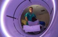 Diagnostic ImagingDiagnostic and interventional scans.
 Anatomical pathology laboratoryAllows to obtain a second opinion from renowned specialists.
 Clinical Analysis LaboratoryComprehensive service in the clinical area.
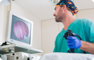 EndoscopyAn accurate diagnosis without conventional surgery.
 ElectrophysiologyFunctional exploration of the central nervous system.
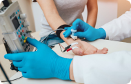 ElectromyographyClinical and neurophysiological evaluation of neuromuscular pathology.
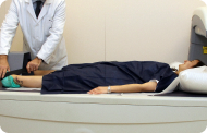 DensitometryDiagnostic technique for checking bone mineral density.
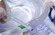 UrodynamicsDiagnosis of urination disorders and incontinence.
- Chequeos médicos

 GeneralUn control inteligente de tu salud
 CompletoUn examen exhaustivo de tu salud
 Completo PlusNuestro chequeo más exclusivo
 ViajerosSi vas a emprender un viaje, tu salud es parte del equipaje
 DeportivoUna revisión a fondo para potenciar tu rendimiento
 CardiológicoUna buena noticia es saber que tu corazón está bajo control
 Para empresasUna herramienta que potencia la satisfacción, productividad y fidelización del empleado
- Diagnostic tests
- Nuestro Centro
- Entorno asistencial Teknon
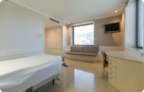 HospitalizaciónHabitaciones luminosas, funcionales y completamente equipadas.
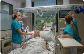 Unidad de SemicríticosDotada de tecnología para diagnósticos y tratamientos que requieren cuidado especial.
 Programa de Alimentación SaludableQueremos mejorar la salud de las personas, por ello promovemos una alimentación saludable, consciente y sostenible en nuestros hospitales.
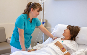 EnfermeríaEquipo de más de 400 profesionales.
 Servicio de Urgencias24 horas al día a tu servicio sin interrupciones.
 Exclusividad / Club TeknonBasado en la alta calidad y el servicio personalizado, ofrecemos un conjunto de servicios que complementan los cuidados médico-asistenciales
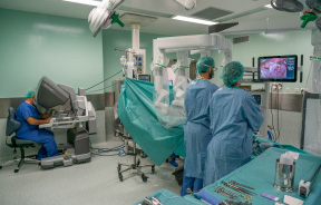 Área QuirúrgicaCuenta con 20 quirófanos, 12 de ellos preparados para cirugía de alto riesgo.
 Programa internacionalUn asistente de este programa le ofrecerá atención integral y personalizada
 Comité de Ética AsistencialAyuda a ciudadanos y profesionales a orientar su actuación en casos de conflictos morales.
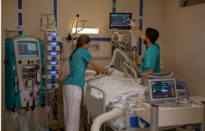 UCI-UCUnidad polivalente con boxes equipados con modernos sistemas de monitorización.
 Atención al pacienteA disposición de todos los pacientes y acompañantes del centro.
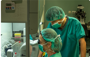 InvestigaciónLa investigación constituye uno de los pilares básicos de Centro Médico Teknon.
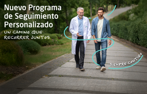 Programa de Seguimiento PersonalizadoTe acompañamos durante tu proceso médico. Organizamos y agendamos tus citas y pruebas.
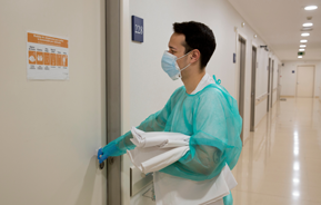 Calidad y Seguridad del PacienteAdoptamos modelos de gestión basados en los estándares más exigentes nacionales e internacionales.
- Entorno asistencial Teknon
- Actualidad
- Actualidad
 NoticiasConoce qué está pasando en Centro Médico Teknon. Consulta nuestra sección de noticias.
 AgendaPuedes encontrar todos los eventos que hemos organizado sobre salud y aquellos temas de actualidad que te pueden interesar. Accede a nuestra agenda de actividades.
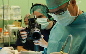 VídeosEn esta sección encontrarás una amplia colección de videos relacionados con nuestras especialidades.
 PodcastTemas médicos de actualidad, tratamientos innovadores, consejos de salud y experiencias de pacientes abordados por nuestros especialistas.
 Contenidos de salud
- Actualidad
- Blog

- Cuadro médico
- Especialidades
- Unidades especializadas
- Instituto del Corazón
- Unidad de Obesidad
- Instituto Oncológico
- Unidad de Medicina y Cirugía sin sangre
- Instituto de Neurociencias
- Unidad de Atención al Lesionado de Tráfico
- Instituto de Neumología
- Unidad de Medicina Marítima
- Instituto de Terapia Regenerativa Tisular
- Unidad de Tratamiento del Dolor
- Clínica del Tenis
- Unidad de Enfermedades Inflamatorias y Autoinmunes
- Unidad de Reproducción
Asistida
- Unidad del Sueño
- Unidad de Síndromes de Sensibilización Central
- Unidades especializadas
- Área diagnóstica
- Diagnostic tests
- Diagnostic ImagingDiagnostic and interventional scans.
- Anatomical pathology laboratoryAllows to obtain a second opinion from renowned specialists.
- Clinical Analysis LaboratoryComprehensive service in the clinical area.
- EndoscopyAn accurate diagnosis without conventional surgery.
- ElectrophysiologyFunctional exploration of the central nervous system.
- ElectromyographyClinical and neurophysiological evaluation of neuromuscular pathology.
- DensitometryDiagnostic technique for checking bone mineral density.
- UrodynamicsDiagnosis of urination disorders and incontinence.
- Chequeos médicos
- GeneralUn control inteligente de tu salud
- CompletoUn examen exhaustivo de tu salud
- Completo PlusNuestro chequeo más exclusivo
- ViajerosSi vas a emprender un viaje, tu salud es parte del equipaje
- DeportivoUna revisión a fondo para potenciar tu rendimiento
- CardiológicoUna buena noticia es saber que tu corazón está bajo control
- Para empresasUna herramienta que potencia la satisfacción, productividad y fidelización del empleado
- Diagnostic tests
- Nuestro Centro
- Entorno asistencial Teknon
- HospitalizaciónHabitaciones luminosas, funcionales y completamente equipadas.
- Unidad de SemicríticosDotada de tecnología para diagnósticos y tratamientos que requieren cuidado especial.
- Programa de Alimentación SaludableQueremos mejorar la salud de las personas, por ello promovemos una alimentación saludable, consciente y sostenible en nuestros hospitales.
- EnfermeríaEquipo de más de 400 profesionales.
- Servicio de Urgencias24 horas al día a tu servicio sin interrupciones.
- Exclusividad / Club TeknonBasado en la alta calidad y el servicio personalizado, ofrecemos un conjunto de servicios que complementan los cuidados médico-asistenciales
- Área QuirúrgicaCuenta con 20 quirófanos, 12 de ellos preparados para cirugía de alto riesgo.
- Programa internacionalUn asistente de este programa le ofrecerá atención integral y personalizada
- Comité de Ética AsistencialAyuda a ciudadanos y profesionales a orientar su actuación en casos de conflictos morales.
- UCI-UCUnidad polivalente con boxes equipados con modernos sistemas de monitorización.
- Atención al pacienteA disposición de todos los pacientes y acompañantes del centro.
- InvestigaciónLa investigación constituye uno de los pilares básicos de Centro Médico Teknon.
- Programa de Seguimiento PersonalizadoTe acompañamos durante tu proceso médico. Organizamos y agendamos tus citas y pruebas.
- Calidad y Seguridad del PacienteAdoptamos modelos de gestión basados en los estándares más exigentes nacionales e internacionales.
- Entorno asistencial Teknon
- Actualidad
- Actualidad
- NoticiasConoce qué está pasando en Centro Médico Teknon. Consulta nuestra sección de noticias.
- AgendaPuedes encontrar todos los eventos que hemos organizado sobre salud y aquellos temas de actualidad que te pueden interesar. Accede a nuestra agenda de actividades.
- VídeosEn esta sección encontrarás una amplia colección de videos relacionados con nuestras especialidades.
- PodcastTemas médicos de actualidad, tratamientos innovadores, consejos de salud y experiencias de pacientes abordados por nuestros especialistas.
- Contenidos de salud
- Actualidad
- Blog

- Unidades especializadas
 Diagnostic tests
- Treatments and Specialities
- Diagnostic Imaging
- Angiography
Angiography
Angiography is a diagnostic technique for examining the vascular system: arteries (arteriography) and veins (phlebography). Angiography can be invasive (a radiological contrast agent is injected through a catheter placed inside the artery or vein, and X-rays are then taken) or non-invasive (CT or MRI angiography, in which the arteries are contrasted by injecting contrast agent intravenously, without the need for catheters). The images obtained provide a detailed map of the vascular system under study (e.g., coronary arteries, cerebral and neck arteries, aorta, lower limb arteries, etc.) and any possible pathology.
In addition to being a diagnostic technique, angiography can also be a therapeutic interventional procedure: once the vascular pathology has been diagnosed, it can be repaired by placing endoprostheses (stents) or intra-arterial inflatable balloons (angioplasties), all without the need for surgery.
Coronary angiography (left heart catheterisation)
Invasive percutaneous diagnostic procedure used to assess the condition of the coronary arteries (morphology, presence or absence of stenosis, degree of severity, etc.).
This technique involves very little risk for the patient and causes minimal discomfort, while the benefits derived from the information it provides are considerable.
The results obtained through coronary angiography are key when deciding on the appropriate treatment for patients with angina pectoris.
How coronary angiography is performed:
- The skin in the groin or on the arm is disinfected.
- Local anaesthesia is applied to this area to insert the catheters into the bloodstream.
- The catheters are advanced through an artery or vein to the heart. This process is monitored under X-ray imaging.
- The catheters reach the origin of the two coronary arteries and, at this point, a radiological contrast agent is injected to ‘fill’ the coronary arteries, obtaining a map of them.
- Multiple radiological images are obtained in different projections, recording the result in a video for analysis.
- The patient may feel temporary warmth from the contrast injection. In some cases, discomfort may occur at the puncture site or a small bruise may appear, which will disappear spontaneously. Arrhythmias, haemorrhages or angina pectoris rarely occur.
 |
 |
|---|---|
| Left coronary artery in right anterior oblique view at 25º. | Right coronary artery in left anterior oblique view at 45º |
Right catheterisation (haemodynamic study)
An invasive percutaneous diagnostic procedure that provides a series of data that cannot be obtained through other diagnostic tests:
- Direct measurements of pressure in the different chambers of the heart
- Measuring the amount of blood pumped
- Observing how blood flows through the heart chambers
This technique involves very little risk for the patient and causes minimal discomfort, while the benefits derived from the information it provides are considerable.
How a haemodynamic study is performed:
- The skin in the groin or on the arm is disinfected.
- Local anaesthesia is applied to this area to insert the catheters into the bloodstream.
- The catheters are advanced through an artery or vein to the heart. This process is monitored under X-ray imaging.
- The pressure in the heart chambers is measured.
- A radiological contrast agent is injected into the left ventricle, which ‘fills’ the chambers, and the process is recorded on video for analysis.
- The patient may feel temporary warmth from the contrast injection. In some cases, discomfort may occur at the puncture site or a small bruise may appear, which will disappear spontaneously. Arrhythmias, haemorrhages or angina pectoris rarely occur.
Electrophysiological study
It is used to accurately diagnose different types of arrhythmias. It also helps guide treatment.
The basics of an electrophysiological study:
The heart is a muscle that pumps blood through a system of vessels, arteries, and veins. The heart has an electrical system that is responsible for emitting the impulses necessary to set the heart rate and adapt it to the body's needs. Disorders of the heart's electrical system cause rhythm disturbances or arrhythmias. Slow arrhythmias occur when the heart rate slows down more than normal, and fast arrhythmias or tachycardias occur when the heart rate speeds up. Electrophysiological study is an invasive percutaneous procedure that diagnoses cardiac arrhythmias and provides guidance on their treatment.
How an electrophysiological study is performed:
- The skin in the groin or on the arm is disinfected.
- Local anaesthesia is applied to this area to insert the catheters into the bloodstream.
- The catheters are advanced through an artery or vein to the heart. This process is monitored under X-ray imaging.
- Catheters are used to record electrical activity, define the type of arrhythmia and locate its source. The electrical activity is displayed on monitors.
- Sometimes it is necessary to administer medication or apply an electric shock. To do this, the patient is anaesthetised.
- The procedure can take anywhere from 30 minutes to several hours.
- When the procedure is complete, the patient must remain at rest for several hours to avoid complications at the puncture site.
Risks associated with this test:
Even if the technique is properly indicated and performed correctly, undesirable effects may occur:
- Minor. It is common for patients to experience palpitations due to the induction of arrhythmias. Sometimes it is necessary to apply an electric shock to resolve a sudden problem.
- Serious. Complications such as phlebitis, venous or arterial thrombosis, or bleeding are rare. Cardiac perforation with cardiac tamponade, or pulmonary or systemic embolism, is very rare. Although these are serious complications that require urgent action.
Ventriculography
An invasive diagnostic test that estimates the size, thickness, and contractile function of the ventricle, as well as providing an accurate assessment of the degree of mitral regurgitation. Cavity visualisation injection requires a large amount of radiological contrast in a short period of time.

Image of the ventricle in diastole and systole
Angioplasty
 Angioplasty is an invasive percutaneous procedure used to ‘open’ coronary arteries that are blocked or narrowed.
A coronary artery stent (vascular endoprosthesis) is a small metal mesh tube that expands inside the artery. A stent is often placed after angioplasty and helps prevent the artery from closing again. A drug-eluting stent (DES) contains medication that helps prevent the artery from closing.
How angioplasty and coronary stenting are performed
- The skin in the groin or on the arm is disinfected.
- Local anaesthesia is applied to this area to insert the catheters into the bloodstream.
- The catheters are advanced through an artery or vein to the heart. This process is monitored under X-ray imaging.
 The catheters reach the origin of the two coronary arteries and, at this point, a radiological contrast agent is injected to ‘fill’ the coronary arteries, obtaining a map of them.
- Once the narrowing (stenosis) of the coronary artery has been located, a catheter with an inflatable balloon is inserted into it to widen the artery.
- Next, the vascular endoprosthesis (stent) can be inserted to keep the artery open.
Percutaneous closure of atrial septal defects (ASD) and percutaneous closure of patent foramen ovale (PFO)
These are percutaneous interventional procedures used to treat congenital heart disease in adults. The procedures prevent blood from flowing from the left atrium to the right atrium. It is performed percutaneously and consists of implanting a double-disc Amplatzer device that self-expands upon implantation, thereby closing the communication between the two discs.

Amplatzer ASD Occluder - Amplatzer PFO Occluder

Amplatzer Occluder placement for ASD