

- Medical directory
- Specialities
- Specialised Units
 Teknon Cardiology Institute
 Obesity Unit
 Teknon Oncology Institute
 Bloodless Medicine and Surgery Unit
 Teknon Institute of Neurosciences
 Traffic Accident Care Unit
 Teknon Pulmonology Unit
 Maritime Medicine Unit
 Institute of Tissue Regenerative Therapy
 Teknon Pain Treatment Unit
 Teknon Tennis Clinic
 Systemic Inflammatory and Autoimmune Diseases Unit
 Assisted Reproduction Unit
 Sleep Unit
 Unidad de Síndromes de Sensibilización Central
- Specialised Units
- Diagnostics
- Diagnostic tests
 Diagnostic ImagingDiagnostic and interventional scans.
 Anatomical pathology laboratoryAllows to obtain a second opinion from renowned specialists.
 Clinical Analysis LaboratoryComprehensive service in the clinical area.
 EndoscopyAn accurate diagnosis without conventional surgery.
 ElectrophysiologyFunctional exploration of the central nervous system.
 ElectromyographyClinical and neurophysiological evaluation of neuromuscular pathology.
 DensitometryDiagnostic technique for checking bone mineral density.
 UrodynamicsDiagnosis of urination disorders and incontinence.
- Medical check-ups

 GeneralThe most intelligent health check-up
 FullA comprehensive examination of your health
 Full PlusOur most exclusive check-up
 TravellersWhen travelling, your health is also part of your luggage
 SportA thorough review to boost your performance
 CardiologyGood news is knowing your heart is under control
 For companiesA tool that enhances employee satisfaction, productivity, and loyalty
- Diagnostic tests
- Our centre
- Teknon Healthcare Service Areas
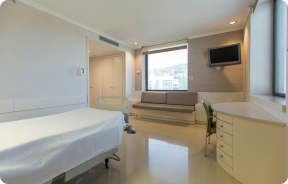 InpatientBright, functional and fully equipped rooms.
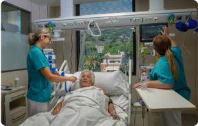 Semi-critical Care UnitEquipped with technology for diagnoses and treatments that require special care.
 Healthy Nutrition ProgrammeWe want to improve people’s health, which is why we promote healthy, conscious and sustainable nutrition at our hospitals.
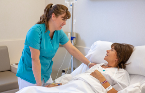 NursingOver 400 professionals.
 Emergency DepartmentUninterrupted operation, 24/7 at your service.
 Exclusivity / Teknon ClubCommitted to superior and individualised service, we furnish a full range of services in conjunction with our medical and healthcare support.
 Surgical AreaA total of twenty (20) operating theatres, 12 of which are equipped for high-risk surgery.
 International ProgrammeA programme agent will provide you with comprehensive, personalised support
 Healthcare Ethics CommitteeGuidance for citizens and professionals in cases of moral conflicts.
 ICU-CCUMultipurpose unit incorporating treatment cubicles equipped with modern monitoring systems.
 Patient servicesAvailable to all our patients and their companions.
 ResearchResearch is one of the cornerstones of Centro Médico Teknon.
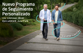 Personalised Follow-up ProgrammeWe accompany you throughout your medical journey. We organise your appointments and tests.
 Patient Quality and SafetyOur management models are based on the most stringent national and international standards.
- Teknon Healthcare Service Areas
- News
- News
 NewsKeep abreast of the events at Centro Médico Teknon. Visit our News section.
 AgendaHere we post upcoming events and discussions on relevant health topics. Visit our Agenda section to see what’s up.
 VideosThis section contains an extensive collection of videos related to our specialities.
 PodcastOur specialists discuss current medical topics, innovative treatments, health advice and patient experience.
 Health content
- News
- Blog

- Medical directory
- Specialities
- Specialised Units
- Teknon Cardiology Institute
- Obesity Unit
- Teknon Oncology Institute
- Bloodless Medicine and Surgery Unit
- Teknon Institute of Neurosciences
- Traffic Accident Care Unit
- Teknon Pulmonology Unit
- Maritime Medicine Unit
- Institute of Tissue Regenerative Therapy
- Teknon Pain Treatment Unit
- Teknon Tennis Clinic
- Systemic Inflammatory and Autoimmune Diseases Unit
- Assisted Reproduction Unit
- Sleep Unit
- Unidad de Síndromes de Sensibilización Central
- Specialised Units
- Diagnostics
- Diagnostic tests
- Diagnostic ImagingDiagnostic and interventional scans.
- Anatomical pathology laboratoryAllows to obtain a second opinion from renowned specialists.
- Clinical Analysis LaboratoryComprehensive service in the clinical area.
- EndoscopyAn accurate diagnosis without conventional surgery.
- ElectrophysiologyFunctional exploration of the central nervous system.
- ElectromyographyClinical and neurophysiological evaluation of neuromuscular pathology.
- DensitometryDiagnostic technique for checking bone mineral density.
- UrodynamicsDiagnosis of urination disorders and incontinence.
- Medical check-ups
- GeneralThe most intelligent health check-up
- FullA comprehensive examination of your health
- Full PlusOur most exclusive check-up
- TravellersWhen travelling, your health is also part of your luggage
- SportA thorough review to boost your performance
- CardiologyGood news is knowing your heart is under control
- For companiesA tool that enhances employee satisfaction, productivity, and loyalty
- Diagnostic tests
- Our centre
- Teknon Healthcare Service Areas
- InpatientBright, functional and fully equipped rooms.
- Semi-critical Care UnitEquipped with technology for diagnoses and treatments that require special care.
- Healthy Nutrition ProgrammeWe want to improve people’s health, which is why we promote healthy, conscious and sustainable nutrition at our hospitals.
- NursingOver 400 professionals.
- Emergency DepartmentUninterrupted operation, 24/7 at your service.
- Exclusivity / Teknon ClubCommitted to superior and individualised service, we furnish a full range of services in conjunction with our medical and healthcare support.
- Surgical AreaA total of twenty (20) operating theatres, 12 of which are equipped for high-risk surgery.
- International ProgrammeA programme agent will provide you with comprehensive, personalised support
- Healthcare Ethics CommitteeGuidance for citizens and professionals in cases of moral conflicts.
- ICU-CCUMultipurpose unit incorporating treatment cubicles equipped with modern monitoring systems.
- Patient servicesAvailable to all our patients and their companions.
- ResearchResearch is one of the cornerstones of Centro Médico Teknon.
- Personalised Follow-up ProgrammeWe accompany you throughout your medical journey. We organise your appointments and tests.
- Patient Quality and SafetyOur management models are based on the most stringent national and international standards.
- Teknon Healthcare Service Areas
- News
- News
- NewsKeep abreast of the events at Centro Médico Teknon. Visit our News section.
- AgendaHere we post upcoming events and discussions on relevant health topics. Visit our Agenda section to see what’s up.
- VideosThis section contains an extensive collection of videos related to our specialities.
- PodcastOur specialists discuss current medical topics, innovative treatments, health advice and patient experience.
- Health content
- News
- Blog


- Specialised Units
 Diagnostic tests
- Treatments and Specialities
- Diagnostic Imaging
- Ultrasound
Ultrasound
Ultrasound (US) is a fast, harmless and dynamic diagnostic imaging method. Using a simple, non-invasive procedure that does not involve radiation, an image of the organs or lesions to be examined is formed. During the scan, the specialist moves the transducer device over a part of the body. This transducer sends sound waves (ultrasound) that ‘bounce’ off the different organs and tissues, returning a new wave that is transformed into a specific image.
- Abdomen
- Abdominal screening
A non-invasive ultrasound technique that provides images of various intra-abdominal organs, such as the liver, gallbladder, spleen, pancreas, kidneys, etc., and assesses whether or not there is disease, determining the extent, type and size of any possible lesions, etc.
- Pelvic screening
A non-invasive, transabdominal ultrasound technique that requires a full bladder in order to visualise the pelvic organs (uterus, ovaries) in relation to the various adjacent internal structures (bladder, intestine, etc.), allowing urinary and gynaecological diseases to be identified and assessed.
- Transrectal screening
Ultrasound technique of the prostate and seminal vesicles performed through the rectum, obtaining images from different planes that allow assessment of the morphology, size and echostructure of the prostate.
- Kidney screening
A non-invasive ultrasound technique that allows us to assess the morphology, size and position of the kidneys. Indicated for assessment of hydronephrosis, lithiasis, study of non-functioning kidneys by urography, evaluation of masses, abscesses and other pathologies.
- Prostate gland screening
A non-invasive transabdominal ultrasound technique that requires proper bladder filling in order to visualise: a) the morphology and size of the prostate and seminal vesicles; b) the walls and interior of the urinary bladder. Indicated for assessing prostate enlargement, inflammatory processes, ruling out bladder tumours, quantifying post-void residual urine, etc.
- Kidney-bladder-prostate screening
A non-invasive transabdominal ultrasound technique that requires proper bladder filling. It allows us to assess: a) the morphology, size and position of the kidneys, b) the walls and interior of the urinary bladder, c) size, prostatic echostructure and seminal vesicles. Indicated for patients with pain, haematuria or urinary tract infection, among other conditions, to rule out inflammatory disease, tumours, prostatic hypertrophy, lithiasis, etc.
- Bladder screening
A non-invasive ultrasound technique that requires adequate filling of the bladder with urine in order to obtain a correct assessment of the walls and interior. Indicated for ruling out tumours, inflammatory processes, lithiasis, megaureters, etc.
- Kidney and bladder
A non-invasive transabdominal ultrasound technique that requires proper bladder filling. It allows us to assess: a) the morphology, size and position of the kidneys b) the walls and interior of the bladder. Indicated for ruling out pathologies in patients with pain, haematuria, urinary tract infection, etc.
- Abdominal and pelvic screening
A non-invasive ultrasound technique used to obtain images of the various intra-abdominal and pelvic organs.
- Abdominal and gynaecological screening
A non-invasive ultrasound technique that provides images of the various intra-abdominal and pelvic organs, including assessment of the uterus and ovaries.
- Transrectal kidney bladder-prostate screening
An ultrasound technique used to obtain images of the kidneys via the abdomen and of the prostate via the rectum.
- Gynaecological screening
- 20-week ultrasound scan
- Ginecològica abdominal
- Parts toves
- Tiroide
Tècnica ultrasonogràfica no invasiva utilitzada per valorar la mida, la morfologia i l'ecoestructura de la glàndula tiroïdal i la detecció de nòduls i processos inflamatoris.
- Paròtida i submaxil·lar
Tècnica ultrasonogràfica no invasiva utilitzada per valorar la mida, la morfologia i l'ecoestructura de les glàndules salivals: paròtide i submaxil·lars, per a la detecció de nòduls, litiasi i processos inflamatoris.
- Coll
Tècnica ultrasonogràfica no invasiva que permet examinar el coll amb la valoració de la morfologia, la mida i l'ecoestructura de les glàndules paròtides, submaxil·lars i tiroides, així com la valoració de possibles ganglis cervicals.
- Paret abdominal
Tècnica ultrasonogràfica no invasiva que permet examinar la paret abdominal anterior, fonamentalment per valorar possibles hèrnies.
- "Bonys"
Tècnica ultrasonogràfica no invasiva que permet examinar qualsevol part de l'anatomia per a la valoració i caracterització de nòduls o tumoracions palpables situades a la pell, al teixit cel·lular subcutani o musculatura.
- Escrotal i testicular
Tècnica ultrasonogràfica no invasiva que permet examinar la bossa escrotal i el seu contingut (testicles, epidídims) amb la valoració d'una possible patologia tumoral, inflamatòria o varicocele (varius testiculars).
- Paret toràcica
Técnica ultrasonográfica, no invasiva, muy útil para valorar la presencia de líquido pleural.
- Muscoloesquelética
- Maluc
Tècnica ultrasonogràfica no invasiva per a la valoració dels tendons de la musculatura glútia, el múscul tensor de la fàscia lata, el trocànter major, el múscul psoasilíac, la inserció dels músculs adductors, el recte anterior i el sartori, els nervis adjacents (nervi femoral i fémoro-cutani lateral), l'articulació coxofemoral i el seu sinovial, per descartar bursitis.
- Genoll
Tècnica ultrasonogràfica no invasiva utilitzada per valorar les articulacions (fémoro-patel·lar i fémoro-tibial), els tendons quadricipitals i rotulians, els lligaments, els meniscs, els tendons distals dels isquitiobials i proximals del tríceps sural, la cintilla iliotibial, la pota d'ànec, nervis adjacents (nervi tibial i nervi peroneal) per descartar bursitis, sinovitis i patologia de ròtula, fèmur i tíbia.
- Turmell i peu
Tècnica ultrasonogràfica no invasiva per a la valoració de les articulacions (tíbia-peroneal distal, tibio-astragalina, les articulacions tarso-metatarsianes metatarsofalàngiques i interfalàngiques), els ossos (tíbia i peroné distal, astràgal, navicular, ossos cuneïformes i cuboides), els tendons (tibial anterior, posterior, flexors i extensors dels dits), els lligaments, la musculatura intrínseca del peu, la fàscia plantar, els nervis adjacents (nervi tibial i nervi sural), descartar bursitis i quists.
- Espatlla
Tècnica ultrasonogràfica no invasiva per a la valoració del braçal rotatori (subescapular, supraespinós i infraespinós), el tendó llarg del bíceps braquial, el tendó del pectoral major, el múscul deltoïdal, l'espai subacromial, les articulacions acromi-clavicular, gleno-humeral, esternoclavicular, el cap humeral, l'acromi, la clavícula, els lligaments, la càpsula articular, el làbrum, els nervis adjacents (nervi supraescapular), descartar quists, bursitis i sinovitis.
- Colze
Tècnica ultrasonogràfica no invasiva usada per a la valoració de les articulacions i el sinovial (húmer-radial, húmer-cubital i radi-cubital proximal), els ossos (húmer distal: epicòndil medial, lateral i olècran, el cap del radi i cúbit proximal), el tendó comú de la musculatura extensora i flexora, el tendó del tríceps braquial, els lligaments per descartar bursitis, sinovitis i patologia nerviosa (nervi mitjà i nervi cubital).
- Canell i mà
Tècnica ultrasonogràfica no invasiva per a la valoració de les articulacions (radicubital distal, radi carpià, cúbit carpià, les articulacions dels cap, carp-metacarpià, metacarpofalàngiques i interfalàngiques), els ossos (cúbit distal, radi distal, escafoides, semilunar, piramidal, pisiforme, trapezi, trapezoide, gran, ganxut, metacarpians i falanges), els tendons i retinacles, els lligaments, la musculatura intrínseca de la mà, nervis adjacents (nervi mitjà i cubital), descartar bursitis i quists.
- Muscular
Ecografia muscular (cuixa, cama, braç, avantbraç, musculatura cervical, dorsal, lumbar, paret toràcica i abdominal). Valoració de lesions traumàtiques, ruptures miotendinoses, patologia nerviosa, descartar col·leccions hemàtiques, Morel-Lavallé i miositis ossificant.
- Eco-doppler
- Puncions evacuants dirigides sota control ecogràfic
Per buidar vessaments articulars, col·leccions hemàtiques, hematomes enquistats, quists o ganglis.
- Inflitracions dirigides sota control ecogràfic
- Mama
- Mama
Tècnica ultrasonogràfica no invasiva que ens permet valorar l'estructura de la glàndula mamària, així com la composició sòlida o líquida dels nòduls mamaris detectats en la mamografia i el seu aspecte benigne o maligne.
- Punció aspirativa (PAAF) de lesions mamàries
Consisteix en la punció per al diagnòstic de lesions nodulars de la mama. Es localitza la lesió sota control ecogràfic, s'insereix una agulla fina a l'interior del nòdul mitjançant aspiració i s'obtenen cèl·lules que es remeten al metge especialista en Citologia (Anatomia Patològica) per a la seva anàlisi.
- Biòpsia amb agulla gruixuda (BAG) de lesions mamàries
Procediment que consisteix en extreure una mostra de teixit mamari amb agulla gruixuda per a l'estudi histològic de tumors de la mama. Prèvia anestèsia local de la zona, se situa l'agulla a la zona que s'ha d'estudiar, sota control ecogràfic i s'obté un cilindre mil·limètric per un sistema de tall automàtic. Posteriorment el metge especialista en Anatomia Patològica farà el diagnòstic de la mostra.
- Marcatge prequirúrgic de lesions mamàries
Procediment que consisteix en col·locar una agulla de morfologia especial "arpó" adjacent a una lesió mamària que posteriorment s'extraurà quirúrgicament. Aquest arpó servei de guia al ginecòleg/cirurgià durant la intervenció, evitant així l'extracció innecessària de teixit mamari sa. Prèvia anestèsia local de la zona, se situa l'agulla a la proximitat del nòdul o lesió que s'ha d'estudiar, sota control ecogràfic.
- Pediàtrica
- Abdominal
Tècnica ultrasonogràfica no invasiva que permet estudiar tots els òrgans abdominal com l'apèndix cecal, el fetge, la vesícula biliar, la melsa, el pàncrees, els intestins i els ronyons, permetent diagnosticar un ventall ampli de patologies, com per exemple: apendicitis aguda, abscessos, traumatismes, tumoracions, etc.
- Renal
Tècnica ultrasonogràfica no invasiva que permet valorar la mida, la morfologia i la situació dels ronyons. S'usa per valorar la dilatació de la pelvis i les calzes renals (ectàsia pièlica, hidronefrosi), masses, malformacions, etc.
- Renal i vesical
Tècnica ultrasonogràfica no invasiva que permet valorar la mida, la morfologia i la situació dels ronyons i la bufeta urinària. Exploració selectiva per a control evolutiu d'hidronefrosi, estudi del ronyó que no funciona per urografia, avaluació de masses, abscessos, megaurèters i altres patologies.
- Malucs
L'ecografia de malucs és actualment l'únic mitjà fiable de diagnòstic abans dels 3 mesos de vida de la displàsia congènita de maluc i que determina el tractament oportú precoçment. També és el mètode més indicat per valorar un vessament líquid articular en la sinovitis transitòria.
- Coll
Tècnica ultrasonogràfica no invasiva que permet valorar les glàndules paròtides, submaxil·lars i el seu aspecte morfològic i ecogràfic en processos inflamatoris i abscessos. També és una tècnica d'elecció per valorar l'aspecte ecogràfic de les adenomegàlies (augment de mida dels ganglis cervicals).
- Ginecològica
Tècnica ultrasonogràfica no invasiva que requereix la bufeta en repleció per visualitzar els òrgans pèlvics (úter i ovaris) i poder valorar la seva mida, ecoestructura, malformacions, formacions quístiques, pubertat precoç, etc.
- Intervencionista