
- Medical directory
- Specialities
- Specialised Units
 Teknon Cardiology Institute
 Obesity Unit
 Teknon Oncology Institute
 Bloodless Medicine and Surgery Unit
 Teknon Institute of Neurosciences
 Traffic Accident Care Unit
 Teknon Pulmonology Unit
 Maritime Medicine Unit
 Institute of Tissue Regenerative Therapy
 Teknon Pain Treatment Unit
 Teknon Tennis Clinic
 Systemic Inflammatory and Autoimmune Diseases Unit
 Assisted Reproduction Unit
 Sleep Unit
 Unidad de Síndromes de Sensibilización Central
- Specialised Units
- Diagnostics
- Diagnostic tests
 Diagnostic ImagingDiagnostic and interventional scans.
 Anatomical pathology laboratoryAllows to obtain a second opinion from renowned specialists.
 Clinical Analysis LaboratoryComprehensive service in the clinical area.
 EndoscopyAn accurate diagnosis without conventional surgery.
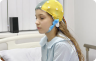 ElectrophysiologyFunctional exploration of the central nervous system.
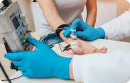 ElectromyographyClinical and neurophysiological evaluation of neuromuscular pathology.
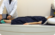 DensitometryDiagnostic technique for checking bone mineral density.
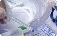 UrodynamicsDiagnosis of urination disorders and incontinence.
- Medical check-ups

 GeneralThe most intelligent health check-up
 FullA comprehensive examination of your health
 Full PlusOur most exclusive check-up
 TravellersWhen travelling, your health is also part of your luggage
 SportA thorough review to boost your performance
 CardiologyGood news is knowing your heart is under control
 For companiesA tool that enhances employee satisfaction, productivity, and loyalty
- Diagnostic tests
- Our centre
- Teknon Healthcare Service Areas
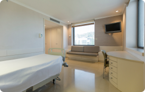 InpatientBright, functional and fully equipped rooms.
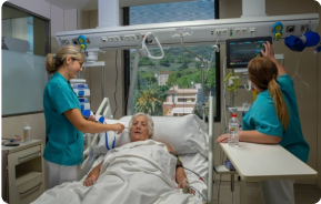 Semi-critical Care UnitEquipped with technology for diagnoses and treatments that require special care.
 Healthy Nutrition ProgrammeWe want to improve people’s health, which is why we promote healthy, conscious and sustainable nutrition at our hospitals.
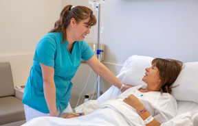 NursingOver 400 professionals.
 Emergency DepartmentUninterrupted operation, 24/7 at your service.
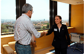 Exclusivity / Teknon ClubCommitted to superior and individualised service, we furnish a full range of services in conjunction with our medical and healthcare support.
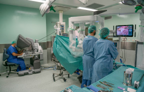 Surgical AreaA total of twenty (20) operating theatres, 12 of which are equipped for high-risk surgery.
 International ProgrammeA programme agent will provide you with comprehensive, personalised support
 Healthcare Ethics CommitteeGuidance for citizens and professionals in cases of moral conflicts.
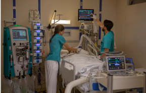 ICU-CCUMultipurpose unit incorporating treatment cubicles equipped with modern monitoring systems.
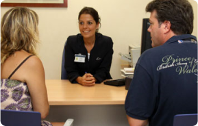 Patient servicesAvailable to all our patients and their companions.
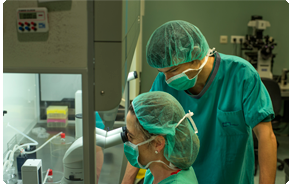 ResearchResearch is one of the cornerstones of Centro Médico Teknon.
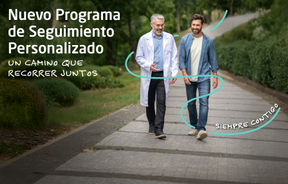 Personalised Follow-up ProgrammeWe accompany you throughout your medical journey. We organise your appointments and tests.
 Patient Quality and SafetyOur management models are based on the most stringent national and international standards.
- Teknon Healthcare Service Areas
- News
- News
 NewsKeep abreast of the events at Centro Médico Teknon. Visit our News section.
 AgendaHere we post upcoming events and discussions on relevant health topics. Visit our Agenda section to see what’s up.
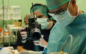 VideosThis section contains an extensive collection of videos related to our specialities.
 PodcastOur specialists discuss current medical topics, innovative treatments, health advice and patient experience.
 Health content
- News
- Blog

- Medical directory
- Specialities
- Specialised Units
- Teknon Cardiology Institute
- Obesity Unit
- Teknon Oncology Institute
- Bloodless Medicine and Surgery Unit
- Teknon Institute of Neurosciences
- Traffic Accident Care Unit
- Teknon Pulmonology Unit
- Maritime Medicine Unit
- Institute of Tissue Regenerative Therapy
- Teknon Pain Treatment Unit
- Teknon Tennis Clinic
- Systemic Inflammatory and Autoimmune Diseases Unit
- Assisted Reproduction Unit
- Sleep Unit
- Unidad de Síndromes de Sensibilización Central
- Specialised Units
- Diagnostics
- Diagnostic tests
- Diagnostic ImagingDiagnostic and interventional scans.
- Anatomical pathology laboratoryAllows to obtain a second opinion from renowned specialists.
- Clinical Analysis LaboratoryComprehensive service in the clinical area.
- EndoscopyAn accurate diagnosis without conventional surgery.
- ElectrophysiologyFunctional exploration of the central nervous system.
- ElectromyographyClinical and neurophysiological evaluation of neuromuscular pathology.
- DensitometryDiagnostic technique for checking bone mineral density.
- UrodynamicsDiagnosis of urination disorders and incontinence.
- Medical check-ups
- GeneralThe most intelligent health check-up
- FullA comprehensive examination of your health
- Full PlusOur most exclusive check-up
- TravellersWhen travelling, your health is also part of your luggage
- SportA thorough review to boost your performance
- CardiologyGood news is knowing your heart is under control
- For companiesA tool that enhances employee satisfaction, productivity, and loyalty
- Diagnostic tests
- Our centre
- Teknon Healthcare Service Areas
- InpatientBright, functional and fully equipped rooms.
- Semi-critical Care UnitEquipped with technology for diagnoses and treatments that require special care.
- Healthy Nutrition ProgrammeWe want to improve people’s health, which is why we promote healthy, conscious and sustainable nutrition at our hospitals.
- NursingOver 400 professionals.
- Emergency DepartmentUninterrupted operation, 24/7 at your service.
- Exclusivity / Teknon ClubCommitted to superior and individualised service, we furnish a full range of services in conjunction with our medical and healthcare support.
- Surgical AreaA total of twenty (20) operating theatres, 12 of which are equipped for high-risk surgery.
- International ProgrammeA programme agent will provide you with comprehensive, personalised support
- Healthcare Ethics CommitteeGuidance for citizens and professionals in cases of moral conflicts.
- ICU-CCUMultipurpose unit incorporating treatment cubicles equipped with modern monitoring systems.
- Patient servicesAvailable to all our patients and their companions.
- ResearchResearch is one of the cornerstones of Centro Médico Teknon.
- Personalised Follow-up ProgrammeWe accompany you throughout your medical journey. We organise your appointments and tests.
- Patient Quality and SafetyOur management models are based on the most stringent national and international standards.
- Teknon Healthcare Service Areas
- News
- News
- NewsKeep abreast of the events at Centro Médico Teknon. Visit our News section.
- AgendaHere we post upcoming events and discussions on relevant health topics. Visit our Agenda section to see what’s up.
- VideosThis section contains an extensive collection of videos related to our specialities.
- PodcastOur specialists discuss current medical topics, innovative treatments, health advice and patient experience.
- Health content
- News
- Blog


- Specialised Units
 Diagnostic tests
- Treatments and Specialities
- Diagnostic Imaging
- Multidetector Computed Tomography
Multidetector Computed Tomography
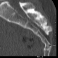
Centro Médico Teknon uses state-of-the-art technology to render three-dimensional reconstruction of the organ under examination. Computed Tomography uses a rotational movement to send successive X-ray emissions to one or more detectors. This data is integrated into a single system which exports it in a ‘volume’ of images representing the scanned region. This provides detailed images with a density resolution far superior to that of conventional radiology.
- Neuroradiology
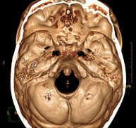
- Skull CT
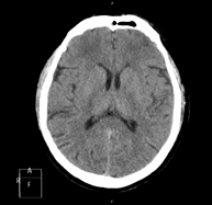
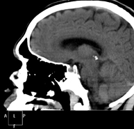
Radiological test that provides high definition anatomical images of the skull (brain stem, cerebellum, cerebrum, cranial calotte, etc.) using CT (Computed Tomography) equipment. Indicated for: trauma, headache, memory disorders, sudden loss of strength in a limb or half of the body.
- Neck CT
Radiological test that provides high definition anatomical images of the neck using CT (Computed Tomography) equipment. Indicated for: thyroid study, control of treated tumours, study of lymph nodes, infections and abscesses.
- Laryngeal CT
Radiological test that provides high definition anatomical images of the larynx using CT (Computed Tomography) equipment. Indicated for: sudden or chronic aphonia, respiratory distress.
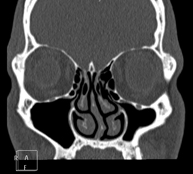
- Orbit CT
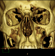
Radiological test that provides high definition anatomical images of the orbits using CT (Computed Tomography) equipment. Indicated for: double vision, infections, trauma, bulging eyes (hyperthyroidism), congenital malformations.
- Pituitary CT
Radiological test that provides high definition anatomical images of the cerebral pituitary gland using CT (Computed Tomography) equipment. Indicated for: suspected pituitary tumour, growth disorder.
- Facial mass CT
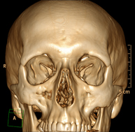
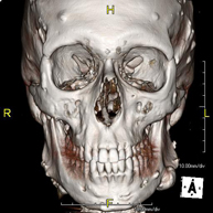
Radiological test that provides high definition anatomical images of the facial mass (face) using CT (Computed Tomography) equipment. Indicated for: tumours, plastic surgery.
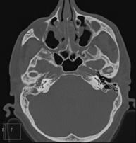
- Ear CT
Radiological test that provides high definition anatomical images of the ear (internal and external auditory canal, eardrum, ossicles of the ear), using CT (Computed Tomography) equipment. Indicated for: hearing disorders, vertigo, dizziness, tinnitus (ringing).
- Dental CT
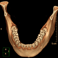
Radiological test that provides high definition anatomical images of the maxillary bone (teeth, dental nerve path) using CT (Computed Tomography) equipment. Indicated for: examination prior to dental extraction, examination prior to implants, tumours, abscess.
- Paranasal Sinuses CT
Radiological test that provides high definition anatomical images of the paranasal sinuses using CT (Computed Tomography) equipment. Indicated for: headache, mucus, facial infections.
- Temporal bone CT
Radiological test that provides high definition anatomical images of the temporal bone (inner, middle and outer ear) using CT (Computed Tomography) equipment. Indicated for: sudden or chronic hearing loss, vertigo, dizziness, congenital malformations.
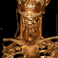
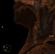
- Angio – Supra-Aortic Trunk CT
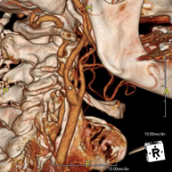
Radiological test that provides high definition anatomical images of the carotid arteries of the neck using CT (Computed Tomography) equipment and the injection of an intravenous contrast agent. The images are then reconstructed in three dimensions (3D). Indicated for: acute cerebral vascular accident, transient vascular accident, carotid bruit.
- Cervical spine CT
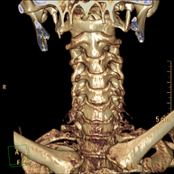
Radiological test that provides high definition anatomical images of the cervical vertebrae using CT (Computed Tomography) equipment. Indicated for: cervical pain without/with irradiation to the arms, trauma.
- Thorax
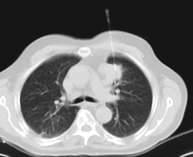
- Chest CT
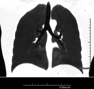
Diagnostic test that provides high definition anatomical images of the chest (lungs, heart, mediastinum, great vessels, rib cage, etc.) using CT (Computed Tomography) equipment. These images are then examined on a workstation that allows bidimensional reconstructions in different planes of space and also 3D reconstructions (volumetric). Some studies require the use of an iodinated contrast agent to improve image definition.
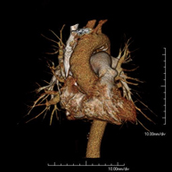
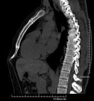
- Thoracic aorta CT angiography
Diagnostic test to examine the thoracic aorta (main artery of the thorax) using CT (Computed Tomography) equipment. This technique requires the use of an iodinated contrast agent, and provides high definition anatomical images. The use of MDCT (Multidetector Computed Tomography) shortens scanning time, reduces radiation dose and improves image quality. The multiple detectors used in certain studies enable imaging to be synchronised with the heartbeat, a technique used to study the aortic valve and aortic root (the first few centimetres), where the heartbeat often distorts images due to movement.
- Pulmonary Artery CT angiography (PTE Study, Pulmonary Thromboembolism)
Diagnostic test to examine the pulmonary arteries using CT (Computed Tomography) equipment to obtain two- and three-dimensional images. This requires the use of an iodinated contrast agent, which will provide improved anatomical definition. This test is mainly indicated in cases of suspected pulmonary thromboembolism (PTE) to rule out or confirm the presence of blood clots inside the arteries.
- High resolution Chest CT
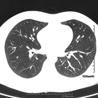
Diagnostic test to examine the lung using CT (Computed Tomography) equipment to obtain two- and three-dimensional images, that allow a highly specific anatomical examination of the lung, being able to assess very small anatomical structures. This technique is very important among patients with suspected lung disease.
- Sternum CT
Utilising an X-ray system and detectors that rotate around a patient, this radiological scan generates images that are computer-reconstructed to facilitate a close examination of the sternum.
- Clavicle CT
Utilising an X-ray system and detectors that rotate around a patient, this radiological scan generates images that are computer-reconstructed to facilitate a close examination of the clavicles.
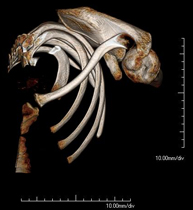
- Ribcage CT
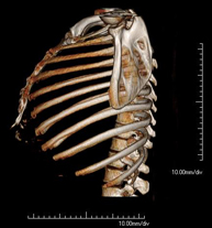
Utilising an X-ray system and detectors that rotate around a patient, this radiological scan generates images that are computer-reconstructed to facilitate a close examination of the ribcage.
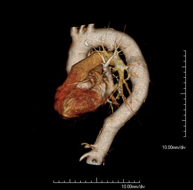
- Coronary CT angiography or Coronary CT
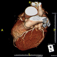
The Coronary CT angiography or non-invasive Coronarography is a diagnostic test to examine arteries of the heart, or coronary arteries, using state-of-the-art MDCT equipment and an iodinated contrast agent to obtain two- and three-dimensional images. Multidetector computed tomography (MDCT) entails high-speed imaging that is beneficial in assessing coronary arteries with high anatomical precision, particularly in evaluating narrowing or stenosis, calcifications, anatomical variants, etc., as its speed prevents the artefact caused by the constant movement of the heart (1,000 images can be obtained in less than 10 seconds). The information obtained requires processing at workstations equipped with specialised software capable of reconstructing the coronary arteries, thereby enabling an assessment to be made of the number, location and characteristics of the lesions. All this information is obtained non-invasively, involving a simple puncture of a peripheral vein (in the arm). To ensure the heart rate stays below 75 bpm, some patients will need preliminary treatment with beta blockers.
- CT-guided thoracic FNA (fine needle aspiration)
This test obtains a sample of tissue from thoracic lesions, such as lung masses, mediastinal masses, bone lesions, etc. This test is performed using local anaesthesia on the puncture area, which is administered with fine-gauge needles. The entire procedure is performed with guidance from images obtained by computed tomography (CT) at various stages of the puncture, using fluoroscopy-CT equipment. After the test, the patient remains in hospital for a few hours. Coagulation tests must be performed before the puncture.
- CT-guided thoracic biopsy

It consists of obtaining a tissue sample from a specific thoracic lesion, such as the lung, mediastinum, sternum, etc. It is sometimes performed under sedation with the help of an anaesthesia team. Needles are used to draw a cylinder sample from the lesion to be studied, which is then sent to the Pathology Department for histological analysis. The entire procedure is performed with guidance from images obtained by computed tomography (CT) at various stages of the biopsy, using fluoroscopy-CT equipment. After the test, the patient remains in hospital under observation. Coagulation tests must be performed before the puncture.
- Thoracic spine CT
Radiological test that involves capturing high-definition anatomical images of the thoracic vertebrae using a CT (computed tomography) scanner. Indicated for: acute/chronic back pain, trauma, spinal misalignment.
- Calcium score

Diagnostic test that measures the amount of calcium that may have accumulated in the atherosclerotic plaques of the coronary arteries using state-of-the-art MDCT equipment. It is a non-invasive test that does not require iodinated contrast. No prior preparation is necessary. Calcified plaques are detected using a specialised workstation that allows the exact amount of calcium to be quantified and given a score, which is known in medical practice as the calcium score.
- Abdomen and pelvis
- Abdomen CT

Diagnostic test that involves obtaining high-definition anatomical images of the abdomen (liver, gallbladder, bile duct, pancreas, spleen, stomach, intestines, kidneys, vascular structures, bladder, uterus and ovaries, etc.) using CT (computed tomography) equipment. These images are then studied at a workstation capable of producing two-dimensional reconstructions in different spatial planes, and also 3D (volumetric) reconstructions. Most studies require the use of iodinated contrast to improve image definition.
- Pelvis CT

Diagnostic test that involves obtaining high-definition anatomical two- and three-dimensional images of the pelvis (bone structures, vascular structures, bladder, uterus and ovaries, prostate and seminal vesicles, ureters, etc.) using CT (computed tomography) equipment. Most studies require the use of iodinated contrast.
- Abdominal and pelvic CT

Diagnostic test that consists of obtaining high-definition anatomical images (bone structures, vascular structures, liver, pancreas, gallbladder, kidneys, adrenal glands, spleen, small and large intestine, bladder, uterus and ovaries, prostate and seminal vesicles, ureters, etc.) using CT (computed tomography) equipment. Most studies require the use of iodinated contrast.
- Liver CT

Diagnostic test that involves obtaining high-definition anatomical two- and three-dimensional images of the liver using CT (computed tomography) equipment. The study must be performed before and after the use of iodinated contrast, carrying it out in different ‘hepatic phases’ to correctly assess all structures: hepatic parenchyma, intra- and extrahepatic bile ducts, gallbladder, hepatic vessels (hepatic artery, portal vein and suprahepatic veins) and adjacent structures (stomach, duodenum, inferior vena cava, pancreatic gland, etc.). This test is particularly useful in the study of liver damage, chronic liver disease, etc.
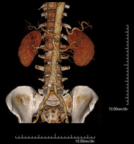
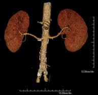
- Kidney CT

Diagnostic test that involves obtaining high-definition anatomical two- and three-dimensional images of the kidney and urinary system using CT (computed tomography) equipment. The study is performed before and after the use of iodinated contrast in different ‘renal phases’ for functional and anatomical assessment (renal parenchyma, ureters, urinary bladder, renal arteries and veins, etc.), as well as adjacent structures (inferior vena cava, abdominal aorta, liver, spleen, etc.). It is particularly recommended when kidney damage is suspected, in patients with blood in their urine or haematuria, etc.
- Urological CT

Diagnostic test that involves obtaining high-definition anatomical two- and three-dimensional images of the kidney and urinary system using CT (computed tomography) equipment. It is particularly recommended for patients suspected of having kidney stones, recurrent urinary tract infections, etc. The test is performed without using iodine contrast (only in certain cases will it be necessary to complete the test with iodine contrast).
- Pancreas CT

Diagnostic test that involves obtaining high-definition anatomical two- and three-dimensional images of the pancreas using CT (computed tomography) equipment. The study is performed before and after the use of iodinated contrast in different ‘pancreatic phases’ in order to assess all structures (pancreatic parenchyma, pancreatic duct or Wirsung duct, biliary-pancreatic junction, common bile duct, pancreatic arteries, splenic artery and vein, duodenum, etc.). It is particularly recommended when pancreatic injury is suspected, in patients with acute or chronic pancreatitis, etc.
- Abdominal aorta CT angiography

A non-invasive diagnostic test that involves studying the abdominal aorta by obtaining high-definition anatomical images using CT (computed tomography) equipment and iodinated contrast. With the aid of workstations specialised for arterial studies, the image quality supports 2D and 3D reconstructions. It is indicated in patients with vascular disease (atherosclerosis), aortic aneurysms, abdominal pain of possible vascular origin, pre-surgical studies of lesions adjacent to the abdominal aorta as a vascular ‘map’, etc. Information obtained non-invasively is indispensable for patients requiring percutaneous or surgical processing. In patients who only require tracking of vascular lesions, this technique is the non-invasive technique of choice, together with MRI angiography.
- Renal artery CT angiography

A non-invasive diagnostic test that involves studying the renal arteries by obtaining high-definition anatomical images using CT (computed tomography) equipment and iodinated contrast. With the aid of workstations specialised for arterial studies, the image quality supports 2D and 3D reconstructions. This test is recommended, for example, in patients suffering from refractory hypertension that does not respond to processing, in patients with kidney damage in order to obtain a pre-surgical ‘vascular’ map, etc.
- Aortoiliac CT angiography

A non-invasive diagnostic test that involves examining the iliac arteries and abdominal aorta, obtaining high-definition anatomical images using CT (computed tomography) equipment and iodinated contrast dye. With the aid of workstations specialised for arterial studies, the image quality supports 2D and 3D reconstructions. This test is particularly recommended as a pre-surgical study (vascular map) prior to percutaneous or surgical interventions on the abdominal aorta, as a complementary study in patients with lower limb ischaemia, etc.
- Virtual colonoscopy
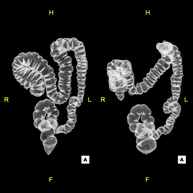

Virtual colonoscopy is a non-invasive technique that allows three-dimensional and two-dimensional visualisation of the large intestine or colon by taking sequential images captured with a state-of-the-art MDCT scanner. The quality of the images allows virtual navigation through the rectum and colon thanks to processing on specialised workstations. Preparation for the test consists of following a low-fibre diet for three days before the test (to cleanse the colon and rectum) and ingesting an iodinated oral contrast agent the day before the test (to mark the stool so that it can be correctly distinguished from any colonic lesions). Unlike fibrocolonoscopy, no sedation or bowel preparation is required. The test is performed in the CT room, where air is blown through a small flexible tube to distend the colon.
- CT-guided abdominal FNA (fine needle aspiration)

It consists in obtaining a tissue sample from a specific lesion located in the abdominal cavity. This test is performed using local anaesthesia on the puncture area, which is administered with fine-gauge needles. The entire procedure is monitored using images obtained by computed tomography (CT) at various stages of the puncture, using fluoroscopy-CT equipment. After the test, the patient remains under observation in hospital for a few hours. Coagulation tests must be performed before the puncture.
- CT-guided abdominal biopsy

It consists in obtaining a tissue sample from a specific lesion located in the abdominal cavity. It is sometimes performed under sedation with the help of an anaesthesia team. Needles are used to draw a cylinder sample from the lesion to be studied, which is then sent to the Pathology Department for histological analysis. The entire procedure is monitored using images obtained by computed tomography (CT) at various stages of the biopsy, using CT fluoroscopy. After the test, the patient remains in hospital under observation. Coagulation tests must be performed before the puncture.
- CT-guided abdominal drainage (abscesses, collections)

It consists of placing a drainage catheter over a collection of fluid located in the abdominal cavity, with the intention of emptying as much of the collection as possible. The patient should keep the drain in place for a few days, usually until it is no longer productive. It is often performed under sedation with the help of an anaesthesia team. The entire procedure is monitored using images obtained by computed tomography (CT) at various stages of the test, using CT fluoroscopy. After the test, the patient remains hospitalised. Coagulation tests must be performed before the test.
- Osteoarticular
- Shoulder CT

Radiological examination based on an X-ray system and detectors that rotate around the patient, reconstructing the images by computer (multidetector computed tomography - MDCT) to study the bones, muscles and joints of the shoulder.
- Elbow CT

Radiological examination based on an X-ray system and detectors that rotate around the patient, reconstructing the images by computer (multidetector computed tomography - MDCT) to study the bones, muscles and joints of the elbow.
- Hand – wrist CT

Radiological examination based on an X-ray system and detectors that rotate around the patient, reconstructing the images by computer (multidetector computed tomography - MDCT) to study the bones, muscles and joints of the hands and wrists.
- Pelvic bone CT

Radiological examination based on an X-ray system and detectors that rotate around the patient, reconstructing the images by computer (multidetector computed tomography - MDCT) to study the bones, muscles and joints of the pelvis.
- Hip CT

Radiological examination based on an X-ray system and detectors that rotate around the patient, reconstructing the images by computer (multidetector computed tomography - MDCT) to study the bones, muscles and joints of the hips.
- Sacroiliac CT

Radiological examination based on an X-ray system and detectors that rotate around the patient, reconstructing the images by computer (multidetector computed tomography - MDCT) to study the sacroiliac joints and rule out inflammatory, traumatic or degenerative diseases.
- Knee CT

Radiological examination based on an X-ray system and detectors that rotate around the patient, reconstructing the images by computer (multidetector computed tomography - MDCT) to study the bones, muscles and joints of the knees.
- Ankle-toe CT

Radiological examination based on an X-ray system and detectors that rotate around the patient, reconstructing the images by computer (multidetector computed tomography - MDCT) to study the bones, muscles and joints of the ankle and foot.
- Lower leg rotational study using CT [patella, tibial tuberosity to trochlear groove (TT-TG) distance]

Radiological examination based on an X-ray system and detectors that rotate around the patient, reconstructing the images by computer (multidetector computed tomography - MDCT) to calculate a series of measurements at the hips, knees and ankles with a view to solving problems affecting the rotation and angulation of the lower limbs.
- TC Huesos largos
 Exploración radiológica que mediante un sistema de rayos X y detectores que giran alrededor del paciente, reconstruyendo las imágenes por ordenador (TC Multidetector), permite el estudio de huesos largos (tibia, peroné, fémur, húmero, radio y cúbito).
 - Biopsia ósea guiada por TC
 Consiste en obtener una muestra de tejido de una determinada lesión ósea. En ocasiones se realiza bajo sedación, con la ayuda del equipo de anestesia. Se utilizan agujas que permiten la obtención de un cilindro de la lesión a estudiar, que se enviará a Anatomía Patológica para su análisis histológico. Todo el procedimiento se realiza controlado con imágenes obtenidas por Tomografía Computarizada (TC) en varios momentos de la biopsia, mediante el empleo de Fluoroscopia-TC. Tras la prueba, el paciente permanece hospitalizado para controlar su evolución. Es necesario aportar pruebas de coagulación antes de la punción.
 - Angio-TC arterial extremidades inferiores
 Prueba diagnóstica no invasiva que consiste en el estudio vascular del sector aorto-ilíaco y de los vasos arteriales de ambas extremidades inferiores obteniendo imágenes de alta definición anatómica mediante el empleo de un equipo de TC Multidetector de última generación y de contraste yodado. La calidad de las imágenes permite realizar reconstrucciones en 2D y 3D gracias a estaciones de trabajo especializadas en el estudio arterial.
- Columna
- TC Columna cervical
 Prueba radiológica que consiste en obtener imágenes de las vértebras cervicales de alta definición anatómica mediante el empleo de un equipo de TC (Tomografía Computarizada). Indicaciones: dolor cervical sin/con irradiación a brazos, traumatismo, malformaciones congénitas.
 - TC Columna dorsal
 Prueba radiológica que consiste en obtener imágenes de las vértebras dorsales de alta definición anatómica mediante el empleo de un equipo de TC (Tomografía Computarizada). Indicaciones: dolor dorsal, estudio de desviaciones de la columna, traumatismo.
 - TC Columna lumbar
 Prueba radiológica que consiste en obtener imágenes de las vértebras lumbares de alta definición anatómica mediante el empleo de un equipo de TC (Tomografía Computarizada). Indicaciones: dolor lumbar sin/con irradiación a piernas, dificultad para caminar, traumatismo.
 - TC Sacro-cóccix
- Pediatría
- TC Cráneo
 Prueba radiológica que consiste en obtener imágenes del cráneo de alta definición anatómica mediante el empleo de un equipo de TC (Tomografía Computarizada). Indicaciones: cefalea, estudio de tumores, traumatismo craneal.
 - TC Cuello
 Prueba radiológica que consiste en obtener imágenes del cuello de alta definición anatómica mediante el empleo de un equipo de TC (Tomografía Computarizada). Indicaciones: infecciones, abscesos, estudio ganglios.
 - TC Tórax
 Prueba diagnóstica que consiste en obtener imágenes del tórax de alta definición anatómica (pulmones, corazón, mediastino, grades vasos, caja torácica, etc.) mediante el empleo de un equipo de TC (Tomografía Computarizada). Dichas imágenes se estudian posteriormente en una estación de trabajo que permite reconstrucciones bidimendionales en diferentes planos del espacio, y también reconstrucciones tridimensionales (3D: volumétricas). Algunos estudios requieren el empleo de contraste yodado para mejorar la definición de las imágenes.
 - TC Abdomen
 Prueba diagnóstica que consiste en obtener imágenes del abdomen de alta definición anatómica (hígado, vesícula biliar, vía biliar, páncreas, bazo, estómago, intestinos, riñones, estructuras vasculares, vejiga, útero y ovarios, etc.) mediante el empleo de un equipo de TC (Tomografía Computarizada). Dichas imágenes se estudian posteriormente en una estación de trabajo que permite reconstrucciones bidimensionales en diferentes planos del espacio, y también reconstrucciones 3D (volumétricas). La mayoría de estudios requieren el empleo de contraste yodado para mejorar la definición de las imágenes.
 - TC Estudio rotacional EEII
Exploración radiológica que mediante un sistema de rayos X y detectores que giran alrededor del paciente, reconstruyendo las imágenes por ordenador, permite calcular una serie de mediciones a nivel de caderas, rodillas y tobillos para solucionar problemas de rotación y angulación de las extremidades inferiores.
- TC Óseo
 Prueba radiológica que consiste en obtener imágenes de los huesos de alta definición anatómica, mediante el empleo de un equipo de TC (Tomografía Computarizada). Indicaciones: traumatismo, estudio de lesiones focales óseas.
 - TC Bajo Sedación
 Exploración radiológica que mediante un sistema de rayos X y detectores que giran alrededor del paciente, reconstruyendo las imágenes por ordenador, permite el estudio de cualquier región del cuerpo. Se realiza bajo sedación, con la colaboración del equipo de Anestesia. En los pacientes pediátricos es de gran ayuda porque permite realizar exploraciones sin falsas imágenes de movimiento (lactantes, niños de corta edad, etc. en los que la situación clínica lo requiera).
- Estudios Vasculares
- Angio-TC-Troncos Supraaórticos
 Prueba radiológica que consiste en obtener imágenes de las arterias carótidas del cuello de alta definición anatómica mediante el empleo de un equipo de TC (Tomografía Computarizada) y la inyección de contraste intravenoso. Posteriormente, las imágenes son reconstruidas en tres dimensiones (3D). Indicaciones: accidente vascular cerebral agudo, accidente vascular transitorio, soplo carotídeo.
 - Angio-TC Aorta torácica
 Prueba diagnóstica que consiste en el estudio de la aorta torácica (principal arteria del tórax) mediante el empleo de un equipo de TC (Tomografía Computarizada). Esta técnica proporciona imágenes de alta definición anatómica. En la mayoría de los casos es necesario el empleo de contraste yodado. El uso del TCMD (TC Multidetector) acorta el tiempo de exploración, disminuye la dosis de radiación y mejora la calidad de imagen. Gracias a los múltiples detectores en determinados estudios se puede acoplar la obtención de la imagen con el latido cardíaco, técnica que permite el estudio de la válvula aórtica y de la raíz de la arteria aorta (primeros centímetros), donde el latido del corazón suele provocar falsas imágenes a causa del movimiento.
 - Angio-TC Arterias pulmonares (Estudio Tep, Tromboembolismo Pulmonar)
 Prueba diagnóstica que consiste en el estudio de las arterias pulmonares mediante el empleo de un equipo de TC (Tomografía Computarizada) obteniendo imágenes bi y tridimensionales. En este estudio es imprescindible el uso de contraste yodado, que permitirá una mejor definición anatómica. Esta prueba está principalmente indicada en los casos de sospecha de tromboembolismo pulmonar (TEP) para descartar o confirmar la presencia de coágulos sanguíneos en el interior de las arterias.
 - Angio-TC Cardíaco
 El angioTC Cardíaco o Coronariografía no invasiva es una prueba diagnóstica que consiste en el estudio de las arterias del corazón o arterias coronarias mediante el empleo de un equipo de TC Multidetector de última generación (64 coronas o filas de detectores) y de contraste yodado, obteniendo imágenes bi y tridimensionales. El TC Multidetector 64 o TCMD64 permite una adquisición de imágenes tan rápida, que se pueden valorar las arterias coronarias con una alta precisión anatómica (estrechamientos o estenosis, calcificaciones, variantes anatómicas, etc.), ya que, gracias a su rapidez, evita el artefacto que provoca el movimiento constante del corazón (tarda menos de diez segundos en adquirir unas 1000 imágenes). La información obtenida precisa de un tratamiento en estaciones de trabajo con programas especializados en la reconstrucción de las arterias coronarias que permiten valorar el número, la localización y las características de las lesiones. Toda esta información se obtiene de manera no invasiva: sólo se requiere la punción de una vena periférica (en el brazo). Es necesario que la frecuencia cardíaca no supere los 75 latidos por minuto, por lo que algunos pacientes deberán realizar un tratamiento previo con un fármaco betabloqueante.
 - Angio-TC aorta abdominal
 Prueba diagnóstica no invasiva que consiste en el estudio de la arteria aorta abdominal obteniendo imágenes de alta definición anatómica mediante el empleo de un equipo de TC (Tomografía Computarizada) y de contraste yodado. La calidad de las imágenes permite realizar reconstrucciones en 2D y 3D gracias a estaciones de trabajo especializadas en el estudio arterial. Está indicado en aquellos pacientes con enfermedad vascular (aterosclerosis), en aneurismas de aorta, en pacientes con dolor abdominal de posible origen vascular, en estudios pre-quirúrgicos de lesiones adyacentes a la aorta abdominal como el "mapa" vascular, etc. La información obtenida de forma no invasiva es indispensable para los pacientes que requieren tratamiento percutáneo o quirúrgico. En aquellos pacientes que solo requieren seguimiento de las lesiones vasculares, esta técnica es la técnica no invasiva de elección junto con la angio-RM.
 - Angio-TC Arterias renales
 Prueba diagnóstica no invasiva que consiste en el estudio de las arterias renales obteniendo imágenes de alta definición anatómica mediante el empleo de un equipo de TC (Tomografía Computarizada) y de contraste yodado. La calidad de las imágenes permite realizar reconstrucciones en 2D y 3D gracias a estaciones de trabajo especializadas en el estudio arterial. Esta prueba está indicada, por ejemplo, en aquellos pacientes que sufren de HTA refractaria al tratamiento, en pacientes con lesiones renales para tener un mapa "vascular" pre-quirúrgico, etc.
 - Angio-TC Aorto-ilíaco
 Prueba diagnóstica no invasiva que consiste en el estudio de las arterias ilíacas y la aorta abdominal obteniendo imágenes de alta definición anatómica mediante el empleo de un equipo de TC (Tomografía Computarizada) y de contraste yodado. La calidad de las imágenes permite realizar reconstrucciones en 2D y 3D gracias a estaciones de trabajo especializadas en el estudio arterial. Esta prueba está especialmente indicada como estudio pre-quirúrgico (mapa vascular) antes de intervenciones percutáneas o quirúrgicas de aorta abdominal, estudio complementario en pacientes con isquemia de miembros inferiores, etc.
 - Angio-TC Arterial extremidades inferiores
 Prueba diagnóstica no invasiva que consiste en el estudio vascular del sector aorto-ilíaco y de los vasos arteriales de ambas extremidades inferiores obteniendo imágenes de alta definición anatómica, mediante el empleo de un equipo de TC Multidetector de última generación y de contraste yodado. La calidad de las imágenes permite realizar reconstrucciones en 2D y 3D gracias a estaciones de trabajo especializadas en el estudio arterial.
- Intervencionismo guiado por TC
- Ablación por radiofrecuencia de tumores óseos
- Colocación de Arpón interpulmonar prequirúrgico
- PAAF (Punción) Tórax guiada por TC
 Prueba que consiste en obtener una muestra de tejido de lesiones torácicas, como por ejemplo masas pulmonares, mediastínicas, lesiones óseas, etc. Para ello se administra anestesia local sobre la zona de la punción, la cual se realiza con agujas de fino calibre. Todo el procedimiento se realiza controlado con imágenes obtenidas por Tomografía Computarizada (TC) en varios momentos de la punción, mediante el empleo de Fluoroscopia-TC. Tras la prueba, el paciente permanece unas horas hospitalizado. Es necesario aportar pruebas de coagulación antes de la punción.
 - Biopsia tórax guiada por TC
 Consiste en obtener una muestra de tejido de una determinada lesión torácica, como por ejemplo del pulmón, del mediastino, del esternón, etc. A veces se realiza bajo sedación, con la ayuda del equipo de anestesia. Se utilizan agujas que permiten la obtención de un cilindro de la lesión a estudiar, que se enviará a Anatomía Patológica para su análisis histológico. Todo el procedimiento se realiza controlado con imágenes obtenidas por Tomografía Computarizada (TC) en varios momentos de la biopsia, mediante el empleo de Fluoroscopia-TC. Tras la prueba, el paciente permanece hospitalizado para controlar su evolución. Es necesario aportar pruebas de coagulación antes de la punción.
 - PAAF (punción) abdominal guiada por TC
 Consiste en obtener una muestra de tejido de una determinada lesión localizada en la cavidad abdominal. Para ello se administra anestesia local sobre la zona de la punción, la cual se realiza con agujas de fino calibre. Todo el procedimiento se realiza controlado con imágenes obtenidas por Tomografía Computarizada (TC) en varios momentos de la punción, mediante el empleo de Fluoroscopia-TC. Tras la prueba, el paciente permanece unas horas hospitalizado para controlar su evolución. Es necesario aportar pruebas de coagulación antes de la prueba.
 - Biopsia abdominal guiada por TC
 Consiste en obtener una muestra de tejido de una determinada lesión localizada en la cavidad abdominal. A veces se realiza bajo sedación, con la ayuda del equipo de anestesia. Se utilizan agujas que permiten la obtención de un cilindro de la lesión a estudiar que se enviará a Anatomía Patológica para su análisis histológico. Todo el procedimiento se realiza controlado con imágenes obtenidas por Tomografía Computarizada (TC) en varios momentos de la biopsia, mediante el empleo de Fluoroscopia-TC. Tras la prueba, el paciente permanece hospitalizado para controlar su evolución. Es necesario aportar pruebas de coagulación antes de la punción.
 - Drenaje abdominal guiado por TC
 Consiste en colocar un catéter de drenaje sobre una colección líquida localizada en la cavidad abdominal, con la intención de vaciar el máximo posible dicha colección. El paciente deberá mantener el drenaje algunos días, normalmente hasta que no sea productivo. A menudo se realiza bajo sedación, con la ayuda del equipo de anestesia. Todo el procedimiento se realiza controlado con imágenes obtenidas por Tomografía Computarizada (TC) en varios momentos de la prueba, mediante el empleo de Fluoroscopia-TC. Tras la prueba, el paciente permanece hospitalizado. Es necesario aportar pruebas de coagulación antes de la prueba.
 - Biopsia ósea guiada por TC
 Exploración invasiva que permite obtener muestras de tejido óseo, principalmente procedente de tumoraciones. El TC es utilizado para seleccionar la zona de punción y guiar las agujas de biopsia hasta la tumoración. Se aplica anestesia local y ocasionalmente sedación. La duración del procedimiento dependerá de la dificultad técnica de este.