

- Quadre mèdic
- Especialitats
- Unitats especialitzades
 Institut del Cor Teknon
 Unitat d’Obesitat
 Institut Oncològic
 Unitat d’Medicina i cirurgia sense sang
 Institut de Neurociències
 Unitat d’atenció al lesionat de trànsit
 Unitat de Pneumologia
 Unitat de Medicina Marítima
 Institut de Teràpia Regenerativa Tissular
 Unitat de Tractament del Dolor
 Clínica del Tennis
 Unitat de malalties inflamatòries i autoimmunes sistèmiques
 Unitat de Reproducció Assistida
 Unitat del Son
 Unitat de Síndromes de Sensibilització Central
- Unitats especialitzades
- Àrea diagnòstica
- Proves diagnòstiques
 Diagnòstic per la ImatgeExploracions diagnòstiques i intervencionistes.
 Laboratori d’anatomia patològicaDisposa d’una segona opinió per part d’especialistes.
 Laboratori d’Anàlisis ClíniquesServei integral a l’àrea clínica.
 EndoscòpiaDiagnòstic precís sense utilitzar la cirurgia.
 ElectrofisiologiaExploració funcional del Sistema Nerviós Central.
 ElectromiografíaAvaluació clínica i neurofisiològica de la patologia neuromuscular.
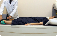 DensitometriaTècnica diagnòstica per comprovar la densitat mineral de l’os.
 UrodinàmiaDiagnòstic dels trastorns de micció i incontinència.
- Revisions mèdiques

 GeneralUn control intel·ligent de la teva salut.
 CompletUn examen exhaustiu de la teva salut.
 Complet PlusLa nostra revisió mèdica més exhaustiva.
 ViatgersSi vols fer un viatge, la teva salut forma part de l’equipatge.
 EsportiuUna revisió a fons per a potenciar el teu rendiment.
 CardiològicUna bona notícia és saber que el teu cor està sota control.
 Per a empresesUna eina que potencia la satisfacció, productivitat i fidelització del treballador.
- Proves diagnòstiques
- El nostre centre
- Entorn assistencial
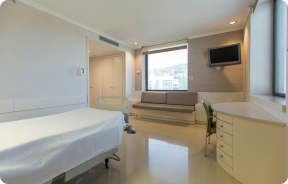 HospitalitzacióAmb habitacions lluminoses, funcionals i completament equipades.
 Unitat de SemicriticsDotada de tecnologia per al diagnòstic i tractament que requereixen una vigilància i cura especial.
 Programa d’Alimentació SaludableVolem millorar la salut de les persones, per això promovem una alimentació saludable, conscient i sostenible als nostres hospitals.
 InfermeriaUn equip de més de 400 professionals.
 Servei d’Urgències24 hores al dia al teu servei sense interrupcions.
 Àrea Pacient PrivatBasat en l’alta qualitat i el servei personalitzat, oferim un conjunt de serveis que complementen les cures mèdico-assistencials
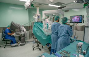 L’Àrea QuirúrgicaDisposa de 20 quiròfans, 12 d’ells preparats per a la cirurgia d’alt risc.
 International programUn assistent d’aquest programa us oferirà atenció integral i personalitzada
 Comitè d’Ètica AssistencialAjuda els ciutadans i als professionals de la salut a orienta la seva actuació en casos de conflictes morals.
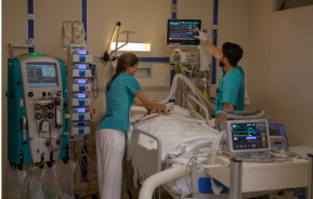 UCI-UCUnitat polivalent que disposa de boxes completament equipats amb el sistema més modern de monitorització.
 Atenció al PacientA disposició de tots els pacients i acompanyants del centre.
 InvestigacióLa investigació constitueix un dels pilars bàsics de Centre Mèdic Teknon.
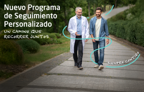 Programa de Seguiment PersonalitzatT’acompanyem durant el teu procés mèdic. Organitzem i agendem les teves cites i proves.
 Qualitat i Seguretat del PacientAdoptem models de gestió basats en els estàndards més exigents nacionals i internacionals.
- Entorn assistencial
- Actualitat
- Actualitat
 NotíciesConeix el que està passant a Centro Médico Teknon. Consulta la nostra secció de notícies.
 AgendaPots trobar tots els esdeveniments que hem organitzat sobre salut i aquells temes d’actualitat que et poden interessar. Accedeix a la nostra agenda d’activitats.
 VídeosEn aquesta secció trobaràs una àmplia col·lecció de vídeos relacionats amb les nostres especialitats.
 PodcastTemes mèdics d’actualitat, tractaments innovadors, consells de salut y experiències de pacients abordabats pels nostres especialistes.
 Continguts de salut
- Actualitat
- Blog

- Quadre mèdic
- Especialitats
- Unitats especialitzades
- Institut del Cor Teknon
- Unitat d’Obesitat
- Institut Oncològic
- Unitat d’Medicina i cirurgia sense sang
- Institut de Neurociències
- Unitat d’atenció al lesionat de trànsit
- Unitat de Pneumologia
- Unitat de Medicina Marítima
- Institut de Teràpia Regenerativa Tissular
- Unitat de Tractament del Dolor
- Clínica del Tennis
- Unitat de malalties inflamatòries i autoimmunes sistèmiques
- Unitat de Reproducció Assistida
- Unitat del Son
- Unitat de Síndromes de Sensibilització Central
- Unitats especialitzades
- Àrea diagnòstica
- Proves diagnòstiques
- Diagnòstic per la ImatgeExploracions diagnòstiques i intervencionistes.
- Laboratori d’anatomia patològicaDisposa d’una segona opinió per part d’especialistes.
- Laboratori d’Anàlisis ClíniquesServei integral a l’àrea clínica.
- EndoscòpiaDiagnòstic precís sense utilitzar la cirurgia.
- ElectrofisiologiaExploració funcional del Sistema Nerviós Central.
- ElectromiografíaAvaluació clínica i neurofisiològica de la patologia neuromuscular.
- DensitometriaTècnica diagnòstica per comprovar la densitat mineral de l’os.
- UrodinàmiaDiagnòstic dels trastorns de micció i incontinència.
- Revisions mèdiques
- GeneralUn control intel·ligent de la teva salut.
- CompletUn examen exhaustiu de la teva salut.
- Complet PlusLa nostra revisió mèdica més exhaustiva.
- ViatgersSi vols fer un viatge, la teva salut forma part de l’equipatge.
- EsportiuUna revisió a fons per a potenciar el teu rendiment.
- CardiològicUna bona notícia és saber que el teu cor està sota control.
- Per a empresesUna eina que potencia la satisfacció, productivitat i fidelització del treballador.
- Proves diagnòstiques
- El nostre centre
- Entorn assistencial
- HospitalitzacióAmb habitacions lluminoses, funcionals i completament equipades.
- Unitat de SemicriticsDotada de tecnologia per al diagnòstic i tractament que requereixen una vigilància i cura especial.
- Programa d’Alimentació SaludableVolem millorar la salut de les persones, per això promovem una alimentació saludable, conscient i sostenible als nostres hospitals.
- InfermeriaUn equip de més de 400 professionals.
- Servei d’Urgències24 hores al dia al teu servei sense interrupcions.
- Àrea Pacient PrivatBasat en l’alta qualitat i el servei personalitzat, oferim un conjunt de serveis que complementen les cures mèdico-assistencials
- L’Àrea QuirúrgicaDisposa de 20 quiròfans, 12 d’ells preparats per a la cirurgia d’alt risc.
- International programUn assistent d’aquest programa us oferirà atenció integral i personalitzada
- Comitè d’Ètica AssistencialAjuda els ciutadans i als professionals de la salut a orienta la seva actuació en casos de conflictes morals.
- UCI-UCUnitat polivalent que disposa de boxes completament equipats amb el sistema més modern de monitorització.
- Atenció al PacientA disposició de tots els pacients i acompanyants del centre.
- InvestigacióLa investigació constitueix un dels pilars bàsics de Centre Mèdic Teknon.
- Programa de Seguiment PersonalitzatT’acompanyem durant el teu procés mèdic. Organitzem i agendem les teves cites i proves.
- Qualitat i Seguretat del PacientAdoptem models de gestió basats en els estàndards més exigents nacionals i internacionals.
- Entorn assistencial
- Actualitat
- Actualitat
- NotíciesConeix el que està passant a Centro Médico Teknon. Consulta la nostra secció de notícies.
- AgendaPots trobar tots els esdeveniments que hem organitzat sobre salut i aquells temes d’actualitat que et poden interessar. Accedeix a la nostra agenda d’activitats.
- VídeosEn aquesta secció trobaràs una àmplia col·lecció de vídeos relacionats amb les nostres especialitats.
- PodcastTemes mèdics d’actualitat, tractaments innovadors, consells de salut y experiències de pacients abordabats pels nostres especialistes.
- Continguts de salut
- Actualitat
- Blog


- Unitats especialitzades
 Proves diagnòstiques
- Tractaments i especialitats
- Diagnòstic per la Imatge
- Tomografia Computaritzada Multidetector
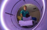
Tomografia Computaritzada Multidetector
Hospital Quirón Teknon disposa d'un mètode d'última generació que permet la reconstrucció tridimensional de l'òrgan estudiat o bé l'obtenció de l'òrgan en els tres plans de l'espai. La Tomografia Computaritzada, mitjançant un moviment de rotació, envia emissions de raigs X successives a un o diversos detectors. Aquestes dades s'integren en un sistema únic que les exporta en un "volum" d'imatges que representen la regió explorada. D'aquesta manera s'obtenen imatges detallades, amb una resolució de densitat molt superior a la de la radiologia convencional.
- Neurorradiologia
- TC Crani
 Prova radiològica que consisteix en obtenir imatges del crani d'alta definició anatòmica (tronc cerebral, cerebel, cervell, calota cranial, etc. ) mitjançant l'ús d'un equip de TC (Tomografia Computeritzada). Indicacions: traumatismes, cefalea, trastorns de la memòria, pèrdua de força sobtada en una extremitat o meitat del cos.
 - TC Coll
 Prova radiològica que consisteix en obtenir imatges del coll d'alta definició anatòmica, mitjançant l'ús d'un equip de TC (Tomografia Computeritzada). Indicacions: estudi de la tiroide, control de tumors tractats, estudi de ganglis, infeccions i abscessos.
 - TC Laringe
 Prova radiològica que consisteix en obtenir imatges de la laringe d'alta definició anatòmica, mitjançant l'ús d'un equip de TC (Tomografia Computeritzada). Indicacions: afonia sobtada o crònica, dificultat respiratòria.
 - TC Òrbites
 Prova radiològica que consisteix en obtenir imatges de les òrbites d'alta definició anatòmica, mitjançant l'ús d'un equip de TC (Tomografia Computeritzada). Indicacions: visió doble, infeccions, traumatismes, ulls sortint (hipertiroidisme), malformacions congènites.
 - TC Hipòfisi
 Prova radiològica que consisteix en obtenir imatges de la hipòfisi cerebral d'alta definició anatòmica, mitjançant l'ús d'un equip de TC (Tomografia Computeritzada). Indicacions: sospita de tumor hipofisari, trastorn del creixement.
 - TC Massís facial

 Prova radiològica que consisteix en obtenir imatges del massís facial (cara) d'alta definició anatòmica, mitjançant l'ús d'un equip de TC (Tomografia Computeritzada). Indicacions: tumors, cirurgia plàstica.
 - TC Oïda
 Prova radiològica que consisteix en obtenir imatges de la oïda d'alta definició anatòmica, (conducte auditiu intern i extern, timpà, ossets de l'oïda) mitjançant l'ús d'un equip de TC (Tomografia Computeritzada). Indicacions: trastorns de l'audició, quadres vertiginosos, mareigs, acúfens (xiulets).
 - TC Dental
 Prova radiològica que consisteix en obtenir imatges dels ossos maxil·lars d'alta definició anatòmica (peces dentals, trajecte del nervi dentari), mitjançant l'ús d'un equip de TC (Tomografia Computeritzada). Indicacions: estudi previ a l'extracció dental, estudi previ als implants, tumors, abscés.
 - TC Sins paranasals
 Prova radiològica que consisteix en obtenir imatges dels sins paranasals d'alta definició anatòmica, mitjançant l'ús d'un equip de TC (Tomografia Computeritzada). Indicacions: cefalea, tos crònica, mucositat, infeccions facials.
 - TC Penyal
 Prova radiològica que consisteix en obtenir imatges de l'os penyal del temporal (oïda interna, mitjana i externa) d'alta definició anatòmica, mitjançant l'ús d'un equip de TC (Tomografia Computeritzada). Indicacions: pèrdua d'audició sobtada o crònica, quadres vertiginosos, mareig, malformacions congènites.
 - Angio-TC Troncs Supraaòrtics
 Prova radiològica que consisteix en obtenir imatges de les artèries caròtides del coll d'alta definició anatòmica, mitjançant l'ús d'un equip de TC (Tomografia Computeritzada) i la injecció de contrast intravenós. Posteriorment, les imatges són reconstruïdes en tres dimensions (3D). Indicacions: accident vascular cerebral agut, accident vascular transitori, buf cardíac.
 - TC Columna cervical
 Prova radiològica que consisteix en obtenir imatges de les cervicals vertebrals d'alta definició anatòmica, mitjançant l'ús d'un equip de TC (Tomografia Computeritzada). Indicacions: cervicàlgia amb/ sense irradiació a braços, traumatisme.
- Tòrax
- TC Tòrax
 Prova diagnòstica que consisteix en obtenir imatges del tòrax d'alta definició anatòmica (pulmons, cor, mediastí, grans vasos, caixa toràcica, etc. ) mitjançant l'ús d'un equip de TC (Tomografia Computeritzada). Aquestes imatges s'estudien posteriorment en una estació de treball que permet reconstruccions bidimensionals en diferents plànols de l'espai i també reconstruccions 3D (volumètriques). Alguns estudis requereixen l'ús de contrast iodat per millorar la definició de les imatges.
 - Angio –TC Aorta toràcica
 Prova diagnòstica que consisteix en l'estudi de l'aorta toràcica (principal artèria del tòrax) mitjançant l'ús d'un equip de TC (Tomografia Computeritzada). Aquesta tècnica requereix l'ús de contrast iodat i proporciona imatges d'alta definició. L'ús del TCMD (TC multidetector) escurça el temps d'exploració, disminueix la dosi de radiació i millora la qualitat d'imatge. Gràcies als múltiples detectors en determinats estudis es pot acoblar l'obtenció de la imatge amb el batec cardíac, tècnica que permet l'estudi de la vàlvula aòrtica i de l'arrel de l'artèria aorta (primers centímetres) on el batec del cor acostuma a provocar múltiples artefactes de moviment.
 - Angio –TC Artèries pulmonars (estudi TEP, Tromboembòlia pulmonar)
 Prova diagnòstica que consisteix en l'estudi de les artèries pulmonars mitjançant l'ús d'un equip de TC (Tomografia Computeritzada) per obtenir imatges bi i tridimensionals. En aquest estudi és imprescindible l'ús de contrast iodat que permet una millor definició anatòmica. Aquesta prova està principalment indicada en els casos de sospita de tromboembòlia pulmonar (TEP) per descartar o confirmar la presència de coàguls sanguinis a l'interior de les artèries.
 - TC Tòrax d'alta resolució
 Prova diagnòstica que consisteix en l'estudi del pulmó mitjançant l'ús d'un equip de TC (Tomografia Computeritzada) per obtenir imatges bi i tridimensional que permeten un estudi anatòmic altament específic del pulmó per poder valorar les estructures anatòmiques de petites dimensions. És una tècnica molt important en l'estudi dels pacients amb sospita de malaltia pulmonar.
 - TC d'estern
 Exploració radiològica que mitjançant un sistema de raigs X i detectors que giren al voltant del pacient i que reconstrueixen les imatges per ordinador, permet l'estudi detallat de l'estern.
 - TC Clavícules
 Exploració radiològica que mitjançant un sistema de raigs X i detectors que giren al voltant del pacient i que reconstrueixen les imatges per ordinador, permet l'estudi detallat de les clavícules.
 - TC Parrilla costal
 Exploració radiològica que mitjançant un sistema de raigs X i detectors que giren al voltant del pacient i que reconstrueixen les imatges per ordinador, permet l'estudi detallat detalla de la Parrilla.
 - Angio- TC Cardíac o TC Cardíac
 L'angio TC Cardíac o Coronariografia no invasiva és una prova diagnòstica que consisteix en l'estudi de les artèries del cor o artèries coronàries mitjançant l'ús d'un equip TC Multidetector d'última generació i de contrast iodat, i l'obtenció d'imatges bi i tridimensionals. El TC Multidetector o TCMD permet una adquisició d'imatges tan ràpida que es poden valorar les artèries coronàries amb alta precisió anatòmica: estrenyiment o estenosi, calcificacions, variants anatòmiques, etc. ja que gràcies a la seva rapidesa evita l'artefacte que provoca el moviment constant del cor (triga menys de deu segons en adquirir unes 1000 imatges). La informació obtinguda precisa un tractament en estacions de treball amb programes especialitzats en la reconstrucció de les artèries coronàries que permeten valorar el nombre, la localització i les característiques de lesions. Tota aquesta informació s'obté de manera no invasiva: només es requereix la punció d'una vena perifèrica (en el braç). És necessari que la freqüència cardíaca no superi els 75 batecs per minut per això alguns pacients hauran de realitzar un tractament previ amb un fàrmac betabloquejant.
 - PAAF (punció) de tòrax guiada per TC
 Prova que consisteix en obtenir una mostra de teixit de lesions toràciques, com per exemple masses pulmonars, mediastíniques, lesions òssies, etc. Per això s'administra anestèsia local sobre la zona de punció, que es realitza amb agulles de calibre fi. Tot el procediment es realitza controlat amb imatges obtingudes per tomografia computaritzada (TC) en diversos moments de la punció, mitjançant un equip de Fluoroscòpia-TC. Després de la prova, el pacient resta unes hores hospitalitzat. És necessari portar proves de coagulació abans de la punció.
 - Biòpsia tòrax guiada per TC
 Consisteix en obtenir una mostra de teixit d'una determinada lesió toràcica, com per exemple del pulmó, del mediastí, de l'estern, etc. A vegades es realitza sota sedació, amb l'ajuda de l'equip d'anestèsia. S'utilitzen agulles que permeten l'obtenció d'un cilindre de la lesió a estudiar que s'enviarà a Anatomia Patològica per a la seva anàlisi histològica. Tot el procediment es realitza controlat amb imatges obtingudes per tomografia computaritzada (TC) en diversos moments de la biòpsia, mitjançant un equip de Fluoroscòpia-TC. Després de la prova, el pacient resta hospitalitzat per controlar la seva evolució. És necessari portar proves de coagulació abans de la punció.
 - TC Columna Dorsal
Prova radiològica que consisteix en obtenir imatges de les vèrtebres dorsals d'alta definició anatòmica, mitjançant l'ús d'un equip de TC (Tomografia Computeritzada). Indicacions: dolor dorsal agut/crònic, traumatisme, columna desviada.
- Score Càlcic
 Prova diagnòstica que consisteix en la mesura de la quantitat de calci que pot haver acumulat a les plaques d'arteriosclerosi de les artèries coronàries mitjançant l'ús d'un equip de TC Multidetector d'última generació. És una prova no invasiva que no requereix contrast iodat. No necessita preparació prèvia. La detecció de les plaques calcificades es realitza en una estació de treball especialitzada que permet la quantificació exacta de la quantitat de calci i ofereix una puntuació que és el que a la pràctica mèdica s'anomena Score Càlcic.
- Abdomen i pelvis
- TC Abdomen
 Prova diagnòstica que consisteix en l'estudi de l'abdomen d'alta definició anatòmica (fetge, vesícula biliar, via biliar, pàncrees, melsa, estómac, intestins, ronyons, estructures vasculars, bufeta, úter i ovaris, etc.) mitjançant l'ús d'un equip de TC (Tomografia Computeritzada). Aquestes imatges s'estudien posteriorment en una estació de treball que permet obtenir reconstruccions bidimensionals en diferents plànols de l'espai i també reconstruccions 3D (volumètriques). La majoria d'estudis requereixen l'ús de contrast iodat per millorar la definició de les imatges.
 - TC Pelvis
 Prova diagnòstica que consisteix en obtenir imatges bi i tridimensionals de la pelvis d'alta definició anatòmica (estructures òssies, estructures vasculars, bufeta, úter i ovaris, pròstata i vesícules seminals, urèters, etc.) mitjançant l'ús d'un equip de TC (Tomografia Computeritzada). La majoria d'estudis requereixen l'ús de contrast iodat.
 - TC Abdominopèlvic
 Prova diagnòstica que consisteix en obtenir imatges bi i tridimensionals de l'abdomen d'alta definició anatòmica (estructures òssies, estructures vasculars, fetge, pàncrees, vesícula biliar, ronyons, glàndules suprarenals, melsa, intestí prim i gros, bufeta, úter i ovaris, pròstata i vesícules seminals, urèters, etc.) mitjançant l'ús d'un equip de TC (Tomografia Computeritzada). La majoria d'estudis requereixen l'ús de contrast iodat.
 - TC Fetge
 Prova diagnòstica que consisteix en obtenir imatges bi i tridimensionals del fetge d'alta definició anatòmica mitjançant l'ús d'un equip de TC (Tomografia Computeritzada). És necessari realitzar l'estudi abans i després de l'ús de contrast iodat, i realitzar l'estudi en diferents "fases hepàtiques" per valorar correctament totes les estructures: parènquima hepàtic, via biliar intra i extrahepàtic, vesícula biliar, vasos hepàtics (artèria hepàtica, vena porta i venes suprahepàtiques) i estructures adjacents (estómac, duodè, vena cava inferior, glàndula pancreàtica, etc.). Aquesta prova està especialment indicada en l'estudi de lesions hepàtiques, estudi d'hepatopaties cròniques, etc.
 - TC Ronyons
 Prova diagnòstica que consisteix en obtenir imatges bi i tridimensionals del ronyó i del sistema urinari d'alta definició anatòmica mitjançant l'ús d'un equip de TC (Tomografia Computeritzada). Es realitza l'estudi abans i després de l'ús de contrast iodat en diferents "fases renals" per a una valoració funcional i anatòmica: parènquima renal, urèters, bufeta urinària, artèries i venes renals, etc. així com les estructures adjacents (vena cava inferior, aorta abdominal, fetge, melsa, etc.). Està especialment indicat quan hi ha sospita de lesions renals, en pacients amb sang a l'orina o hematúria, etc.
 - TC Urològic
 Prova diagnòstica que consisteix en obtenir imatges bi i tridimensionals del ronyó i del sistema urinari d'alta definició anatòmica mitjançant l'ús d'un equip de TC (Tomografia Computeritzada). Està especialment indicat en aquells pacients en els quals se sospita que hi ha pedres al ronyó, infeccions urinàries de repetició. L'estudi es realitza sense utilitzar contrast iodat (només es casos determinats serà necessari completar l'estudi amb contrast iodat).
 - TC Pàncrees
 Prova diagnòstica que consisteix en obtenir imatges bi i tridimensionals del pàncrees d'alta definició anatòmica mitjançant l'ús d'un equip de TC (Tomografia Computeritzada). Es realitza l'estudi abans i després de l'ús de contrast iodat en diferents "fases pancreàtiques" per poder valorar totes les estructures: parènquima pancreàtic, conducte pancreàtic o de Wirsung, unió bilio-pancreàtica, colèdoc, artèries pancreàtiques, artèria i vena esplèniques, duodè. Està especialment indicant quan hi ha sospita de lesió pancreàtica, en pacients amb pancreatitis aguda o crònica, etc.
 - Angio-TC Aorta abdominal

 Prova diagnòstica no invasiva que consisteix en l'estudi de l'artèria aorta abdominal a través de l'obtenció d'imatges d'alta definició anatòmica mitjançant l'ús d'un equip de TC (Tomografia Computeritzada) i de contrast iodat. La qualitat de les imatges permet realitzar reconstruccions en 2D i 3D gràcies a estacions de treball especialitzades en l'estudi arterial. Està indicat en aquells pacients amb malaltia vascular (arteriosclerosi), en aneurismes d'aorta, en pacients amb dolor abdominal de possible origen vascular, en estudis prequirúrgics de lesions adjacents a l'aorta abdominal com a "mapa" vascular. La informació obtinguda de manera no invasiva és indispensable per als pacients que requereixen tractament percutani o quirúrgic. En aquells pacients que només requereixen seguiment de les lesions vasculars, aquesta tècnica és la tècnica no invasiva d'elecció juntament amb l'angio-RM.
 - Angio-TC Artèries renals

 Prova diagnòstica no invasiva que consisteix en l'estudi de les artèries renals a través de l'obtenció d'imatges d'alta definició anatòmica mitjançant l'ús d'un equip de TC (Tomografia Computeritzada) i de contrast iodat. La qualitat de les imatges permet realitzar reconstruccions en 2D i 3D gràcies a estacions de treball especialitzades en l'estudi arterial. Està indicat en aquells pacients amb malaltia vascular (arteriosclerosi), en aneurismes d'aorta, en pacients amb dolor abdominal de possible origen vascular, en estudis prequirúrgics de lesions adjacents a l'aorta abdominal com a "mapa" vascular. La informació obtinguda de manera no invasiva és indispensable per als pacients que requereixen tractament percutani o quirúrgic. En aquells pacients que només requereixen seguiment de les lesions vasculars, aquesta tècnica és la tècnica no invasiva d'elecció juntament amb l'angio-RM.
 - Angio-TC Aorto-ilíac
 Prova diagnòstica no invasiva que consisteix en l'estudi de les artèries ilíaques i l'aorta abdominal a través de l'obtenció d'imatges d'alta definició anatòmica mitjançant l'ús d'un equip de TC (Tomografia Computeritzada) i de contrast iodat. La qualitat de les imatges permet realitzar reconstruccions en 2D i 3D gràcies a estacions de treball especialitzades en l'estudi arterial. Aquesta prova està especialment indicada com estudi prequirúrgic (mapa vascular) abans d'intervencions percutànies o quirúrgiques d'aorta abdominal, com l'estudi complementari en pacients amb isquèmia de membres inferiors, etc.
 - Colonoscòpia virtual

 La colonoscòpia virtual és una tècnica no invasiva que permet la visualització tridimensional i bidimensional de l'intestí gros o del còlon mitjançant la presa seqüencial d'imatges captades amb TC Multidetector d'última generació. La qualitat de les imatges permet la navegació virtual per l'interior del recte i del còlon gràcies al processament en estacions de treball especialitzades. La preparació de la prova consisteix en realitzar una dieta baixa en fibra tres dies abans de la prova (per netejar el còlon i el recte) i de la ingesta de contrast oral iodat el dia abans de la prova (per marcar les femtes i poder distingir-les correctament de les possibles lesions colòniques). A diferència de la fibrocolonoscòpia, no requereix sedació ni solucions evacuants. La prova es realitza a la sala del TC, on, a través d'un petit tub flexible, s'insufla aire per distendre el còlon.
 - PAAF (Punció) Abdominal guiada per TC
 Consisteix en obtenir una mostra de teixit d'una determinada lesió localitzada a la cavitat abdominal. Per això s'administra anestèsia local sobre la zona de punció, que es realitza amb agulles de calibre fi. Tot el procediment es realitza controlat per imatges obtingudes per Tomografia Computaritzada (TC) en diversos moments de la punció, mitjançant l'ús d'un equip de Fluroscòpia-TC. Després de la prova, el pacient resta unes hores hospitalitzat per controlar la seva evolució. És necessari que portin proves de coagulació abans de la punció.
 - Biòpsia abdominal guiada per TC
 Consisteix en obtenir una mostra de teixit d'una determinada lesió localitzada a la cavitat abdominal. En ocasions es realitza sota sedació, amb l'ajuda de l'equip d'anestèsia. S'utilitzen agulles que permeten l'obtenció d'un cilindre de la lesió que s'ha d'estudiar, que s'enviarà a Anatomia Patològica per a la seva anàlisi histològica. Tot el procediment es realitza controlat per imatges obtingudes per Tomografia Computaritzada (TC) en diversos moments de la biòpsia mitjançant l'ús de Fluoroscòpia –TC. Després de la prova, el pacient resta hospitalitzat per controlar la seva evolució. És necessari que portin proves de coagulació abans de la punció.
 - Drenatge abdominal guiat per TC (abscessos, col·leccions)
 Consisteix en col·locar un catèter de drenatge sobre una col·lecció líquida localitzada a la cavitat abdominal, amb la intenció de buidar el màxim possible aquesta col·lecció. El pacient ha de mantenir el drenatge alguns dies, normalment fins que no sigui productiu. Sovint es realitza sota sedació, amb l'ajuda de l'equip d'anestèsia. Tot el procediment es realitza controlat per imatges obtingudes per Tomografia Computaritzada (TC) en diversos moments de la prova mitjançant l'ús de Fluoroscòpia –TC. Després de la prova, el pacient resta hospitalitzat. És necessari que porti les proves de coagulació abans de la prova.
- Osteoarticular
- TC d'espatlla
 Exploració radiològica que mitjançant un sistema de raigs X i detectors que giren al voltant del pacient i que reconstrueixen les imatges per ordinador (TC Multidetector), permet l'estudi detallat dels ossos, els músculs i les articulacions de l'espatlla.
 - TC de colze
 Exploració radiològica que mitjançant un sistema de raigs X i detectors que giren al voltant del pacient i que reconstrueixen les imatges per ordinador (TC Multidetector), permet l'estudi detallat dels ossos, els músculs i les articulacions del colze.
 - TC de la mà
 Exploració radiològica que mitjançant un sistema de raigs X i detectors que giren al voltant del pacient i que reconstrueixen les imatges per ordinador (TC Multidetector), permet l'estudi detallat dels ossos, els músculs i les articulacions de la mà i el canell.
 - TC de la pelvis òssia
 Exploració radiològica que mitjançant un sistema de raigs X i detectors que giren al voltant del pacient i que reconstrueixen les imatges per ordinador (TC Multidetector), permet l'estudi detallat dels ossos, els músculs i les articulacions de la pelvis.
 - TC de malucs
 Exploració radiològica que mitjançant un sistema de raigs X i detectors que giren al voltant del pacient i que reconstrueixen les imatges per ordinador (TC Multidetector), permet l'estudi detallat dels ossos, els músculs i les articulacions del maluc.
 - TC de sacroilíaques
 Exploració radiològica que mitjançant un sistema de raigs X i detectors que giren al voltant del pacient i que reconstrueixen les imatges per ordinador (TC Multidetector), permet l'estudi detallat de les articulacions sacroilíaques i descartar malalties inflamatòries, traumàtiques o degeneratives.
 - TC de genoll
 Exploració radiològica que mitjançant un sistema de raigs X i detectors que giren al voltant del pacient i que reconstrueixen les imatges per ordinador (TC Multidetector), permet l'estudi detallat dels ossos, els músculs i les articulacions del genoll.
 - TC de turmell-peu
 Exploració radiològica que mitjançant un sistema de raigs X i detectors que giren al voltant del pacient i que reconstrueixen les imatges per ordinador (TC Multidetector), permet l'estudi detallat dels ossos, els músculs i les articulacions del turmell i el peu.
 - TC d'estudi rotacional EEII (Bàscula rotuliana, distància TA-GT)
 Exploració radiològica que mitjançant un sistema de raigs X i detectors que giren al voltant del pacient i que reconstrueixen les imatges per ordinador (TC Multidetector), permet calcular una sèrie de mesures a nivell de maluc, genolls i turmells per solucionar problemes de rotació i angulació de les extremitats inferiors.
 - TC d'ossos llargs
 Exploració radiològica que mitjançant un sistema de raigs X i detectors que giren al voltant del pacient i que reconstrueixen les imatges per ordinador (TC Multidetector), permet l'estudi detallat dels ossos llargs (tíbia, peroné, fèmur, húmer, radi i cúbit).
 - Biòpsia òssia guiada per TC
 Consisteix en obtenir una mostra de teixit d'una determinada lesió òssia. En ocasions es realitza sota sedació, amb l'ajuda de l'equip d'anestèsia. S'utilitzen agulles que permeten l'obtenció d'un cilindre de la lesió que s'ha d'estudiar, que s'enviarà a Anatomia Patològica per a la seva anàlisi histològica. Tot el procediment es realitza controlat amb imatges obtingudes per Tomografia Computaritzada (TC). Després de la prova, el pacient resta hospitalitzat per controlar la seva evolució. És necessari que porti les proves de coagulació abans de la punció.
 - Angio-TC arterial d'extremitats inferiors
 Prova diagnòstica no invasiva que consisteix en l'estudi vascular del sector aorto-ilíac i dels vasos arterials d'ambdues extremitats inferiors i l'obtenció d'imatges d'alta definició anatòmica mitjançant l'ús d'un equip de TC Multidetector d'última generació i de contrast iodat. La qualitat de les imatges permet realitzar reconstruccions en 2D i 3D gràcies a estacions de treball especialitzades en l'estudi arterial.
- Columna
- TC Columna cervical
 Prova radiològica que consisteix en obtenir imatges de les vèrtebres cervicals d'alta definició anatòmica, mitjançant l'ús d'un equip de TC (Tomografia Computeritzada). Indicacions: dolor cervical amb/sense irradiació als braços, traumatisme, malformacions congènites.
 - TC Columna dorsal
 Prova radiològica que consisteix en obtenir imatges de les vèrtebres dorsals d'alta definició anatòmica, mitjançant l'ús d'un equip de TC (Tomografia Computaritzada). Indicacions: dolor dorsal, estudi de desviacions de la columna, traumatisme.
 - TC Columna lumbar
 Prova radiològica que consisteix en obtenir imatges de les vèrtebres lumbars d'alta definició anatòmica, mitjançant l'ús d'un equip de TC (Tomografia Computaritzada). Indicacions: dolor lumbar sense / amb irradiació a les cames, dificultat per caminar, traumatisme.
 - TC de sacre-còccix
- Pediatria
- TC de crani
 Prova radiològica que consisteix en obtenir imatges del crani d'alta definició anatòmica, mitjançant l'ús d'un equip de TC (Tomografia Computeritzada). Indicacions: cefalea, estudi de tumors, traumatisme cranial.
 - TC de coll
 Prova radiològica que consisteix en obtenir imatges del coll d'alta definició anatòmica, mitjançant l'ús d'un equip de TC (Tomografia Computeritzada).
 - TC Tòrax
 Prova diagnòstica que consisteix en obtenir imatges del tòrax d'alta definició anatòmica (pulmons, cor, mediastí, grans vasos, caixa toràcica, etc.) mitjançant l'ús d'un equip de TC (Tomografia Computeritzada). Aquestes imatges s'estudien posteriorment en una estació de treball que permet reconstruccions bidimensionals en diferents plans de l'espai i també reconstruccions tridimensionals (3D: volumètriques). Alguns estudis requereixen l'ús de contrast iodat per millorar la definició de les imatges.
 - TC Abdomen
 Prova diagnòstica que consisteix en obtenir imatges de l'abdomen d'alta definició anatòmica (fetge, vesícula biliar, via biliar, pàncrees, melsa, estómac, intestins, ronyons, estructures vasculars, bufeta, úter i ovaris, etc.) mitjançant l'ús d'un equip de TC (Tomografia Computeritzada). Aquestes imatges s'estudien posteriorment en una estació de treball que permet reconstruccions bidimensional en diferents plans de l'espai, i també reconstruccions tridimensionals (3D: volumètriques). La majoria d'estudis requereixen l'ús de contrast iodat per millorar la definició de les imatges.
 - TC d'estudi rotacional EEII
Exploració radiològica que mitjançant un sistema de raigs X i detectors que giren al voltant del pacient i que reconstrueixen les imatges per ordinador, permet calcular una sèrie de mesures a nivell de maluc, genolls i turmells per solucionar problemes de rotació i d'angular de les extremitats inferiors.
- TC Ossi
 Prova radiològica que consisteix en obtenir imatges dels ossos d'alta definició anatòmica, mitjançant l'ús d'un equip de TC (Tomografia Computeritzada). Indicacions: traumatisme, estudi de lesions focals òssies.
 - TC sota sedació
 Exploració radiològica que mitjançant un sistema de raigs X i de detectors que giren al voltant del pacient i que reconstrueixen les imatges per ordinador, permet l'estudi de qualsevol regió del cos. Es realitza sota sedació, amb la col·laboració de l'equip d'Anestèsia. En els pacients pediàtrics és de gran ajuda perquè permet realitzar exploracions sense falses imatges de moviment (lactants, nens de poca edat, etc. i en aquells que la situació clínica ho requereixi).
- Estudis Vasculars
- Angio TC de troncs supraaòrtics
 Prova radiològica que consisteix en obtenir imatges de les artèries caròtides del coll d'alta definició anatòmica, mitjançant l'ús d'un equip de TC (Tomografia Computeritzada) i la injecció de contrast intravenós. Posteriorment, es reconstrueixen les imatges en tres dimensions (3D). Indicacions: accident vascular cerebral agut, accident vascular transitori, buf cardíac.
 - Angio TC- aorta toràcica
 Prova diagnòstica que consisteix en l'estudi de l'aorta toràcica (principal artèria del tòrax) mitjançant l'ús d'un equip de TC (Tomografia Computeritzada). Aquesta tècnica proporciona imatges d'alta definició anatòmica. En la majoria de casos és necessari l'ús de contrast iodat. L'ús del TCMD (TC Multidetector) escurça el temps d'exploració, disminueix la dosi de radiació i millora la qualitat d'imatge. Gràcies als múltiples detectors en determinats estudis es pot afegir l'obtenció de la imatge amb el batec cardíac, tècnica que permet l'estudi de la vàlvula aòrtica i de l'arrel de l'artèria aorta (primers centímetres), on el batec del cor acostuma a provocar falses imatges a causa del moviment.
 - Angio TC d'artèries pulmonars (estudi Tep, tromboembolisme)
 Prova diagnòstica que consisteix en l'estudi de les artèries pulmonars mitjançant l'ús d'un equip de TC (Tomografia Computaritzada) i amb l'obtenció d'imatges bi i tridimensionals. En aquest estudis és imprescindible l'ús de contrast iodat, que permet una millor definició anatòmica. Aquesta prova està principalment indicada en els casos en els quals hi hagi sospita de tromboembolisme pulmonar (TEP) per descartar o confirmar la presència de coàguls sanguinis a l'interior de les artèries.
 - Angio TC cardíac
 L'Angio TC cardíac o la coronariografia no invasiva és una prova diagnòstica que consisteix en l'estudi de les artèries del cor o artèries coronàries mitjançant l'ús d'un equip de TC Multidetector d'última generació (64 corones o files de detectors) i de contrast iodat amb l'obtenció d'imatges bi i tridimensionals. El TC Multidetector 64 o TCMD64 permet una adquisició d'imatges tan ràpida que es poden valorar les artèries coronàries amb alta precisió anatòmica (estrenyiments o estenosis, calcificacions, variants anatòmiques, etc.), ja que, gràcies a la seva rapidesa, evita les falses imatges que provoca el moviment constant del cor (triga menys de deu segons en adquirir unes 1000 imatges). La informació obtinguda d'un tractament en estacions de treball amb programes especialitzats en la reconstrucció de les artèries coronàries que permeten valorar el nombre, la localització i les característiques de les lesions. Tota aquesta informació s'obté de manera no invasiva: només es necessita la punció d'una vena perifèrica (en el braç). És necessari que la freqüència cardíaca no superi els 75 batecs per minut, per això els pacients han de realitzar un tractament previ amb un fàrmac betabloquejant.
 - Angio TC d'aorta abdominal
 Prova diagnòstica no invasiva que consisteix en l'estudi de l'artèria aorta abdominal amb l'obtenció d'imatges d'alta definició anatòmica mitjançant l'ús d'un equip de TC (Tomografia Computaritzada) i contrast iodat. La qualitat de les imatges permet realitzar reconstruccions en 2D i 3D gràcies a estacions de treball especialitzades en l'estudi arterial. Està indicat en aquells pacients que pateixen malaltia vascular (arteriosclerosi), aneurismes d'aorta, en pacient amb dolor abdominal d'un possible origen vascular, en estudis prequirúrgics de lesions adjacents a l'aorta abdominal com el "mapa" vascular, etc. La informació obtinguda de forma no invasiva és indispensable per als pacients que requereixen tractament percutani o quirúrgic. En aquells pacients que només requereixen un seguiment de les lesions vasculars, aquesta tècnica és la tècnica no invasiva d'elecció juntament amb l'angio RM.
 - Angio TC d'artèries renals
 Prova diagnòstica no invasiva que consisteix en l'estudi de les artèries renals amb l'obtenció d'imatges d'alta definició anatòmica mitjançant l'ús d'un equip de TC (Tomografia Computaritzada) i contrast iodat. La qualitat de les imatges permet realitzar reconstruccions en 2D i 3D gràcies a estacions de treball especialitzades en l'estudi arterial. Aquesta prova està indicat en aquells pacients que pateixen HTA refractària al tractament, en pacients amb lesions renals que tenen un mapa vascular prequirúrgic, etc.
 - Angio TC d'aorta-ilíaca
 Prova diagnòstica no invasiva que consisteix en l'estudi de les artèries ilíaques i l'aorta abdominal amb l'obtenció d'imatges d'alta definició anatòmica mitjançant l'ús d'un equip de TC (Tomografia Computaritzada) i contrast iodat. La qualitat de les imatges permet realitzar reconstruccions en 2D i 3D gràcies a estacions de treball especialitzades en l'estudi arterial. Aquesta prova està especialment indicada com a estudi prequirúrgic (mapa vascular) abans d'intervencions percutànies o quirúrgiques d'aorta abdominal, estudi complementari en pacients amb isquèmia de membres inferiors, etc.
 - Angio TC arterial d'extremitats inferiors
 Prova diagnòstica no invasiva que consisteix en l'estudi de vascular del sector aorto-ílíac i dels vasos arterials d'ambdues extremitats inferiors amb l'obtenció d'imatges d'alta definició anatòmica mitjançant l'ús d'un equip de TC (Tomografia Computaritzada) i contrast iodat. La qualitat de les imatges permet realitzar reconstruccions en 2D i 3D gràcies a estacions de treball especialitzades en l'estudi arterial.
- Intervencions guiades per TC
- Ablació per radiofreqüència de tumors ossis
- Col·locació d'Arpó interpulmonar prequirúrgic
- PAAF (Punció) de tòrax guiada per TC
 Prova que consisteix en obtenir una mostra de teixit de lesions toràciques, com per exemple masses pulmonars, mediastíniques, lesions òssies, etc. Per a tal s'administra anestèsia local sobre la zona de punció, que es realitza amb agulles de calibre fi. Tot el procediment es realitza controlat per imatges obtingudes per Tomografia Computaritzada (TC) en diversos moments de la punció mitjançant l'ús de Fluoroscòpia- TC. Després de la prova, el pacient resta unes hores hospitalitzat. És necessari que porti les proves de coagulació abans de la punció.
 - Biòpsia de tòrax guiada per TC
 Consisteix en obtenir una mostra de teixit d'una determinada lesió toràcica, com per exemple del pulmó, del mediastí, de l'estern, etc. A vegades es realitza sota sedació, amb l'ajuda de l'equip d'anestèsia. S'utilitzen agulles que permeten l'obtenció d'un cilindre de la lesió que s'ha d'estudiar, que s'enviarà a Anatomia Patològica per a la seva anàlisi histològica. Tot el procediment es realitza controlat per imatges obtingudes per Tomografia Computaritzada (TC) en diversos moments de la biòpsia mitjançant l'ús de Fluoroscòpia-TC. Després de la prova, el pacient resta hospitalitzat per controlar la seva evolució. És necessari que porti proves de coagulació abans de la punció.
 - PAAF (Punció) abdominal guiada per TC
 Prova que consisteix en obtenir una mostra de teixit de lesions toràciques, com per exemple masses pulmonars, mediastíniques, lesions òssies, etc. Per a tal s'administra anestèsia local sobre la zona de punció, que es realitza amb agulles de calibre fi. Tot el procediment es realitza controlat per imatges obtingudes per Tomografia Computaritzada (TC) en diversos moments de la punció mitjançant l'ús de Fluoroscòpia- TC. Després de la prova, el pacient resta unes hores hospitalitzat. És necessari que porti les proves de coagulació abans de la punció.
 - Biòpsia abdominal guiada per TC
 Consisteix en obtenir una mostra de teixit d'una determinada lesió determinada de lesió localitzada en la cavitat abdominal. A vegades es realitza sota sedació, amb l'ajuda de l'equip d'anestèsia. S'utilitzen agulles que permeten l'obtenció d'un cilindre de la lesió que s'ha d'estudiar, que s'enviarà a Anatomia Patològica per a la seva anàlisi histològica. Tot el procediment es realitza controlat per imatges obtingudes per Tomografia Computaritzada (TC) en diversos moments de la biòpsia mitjançant l'ús de Fluoroscòpia-TC. Després de la prova, el pacient resta hospitalitzat per controlar la seva evolució. És necessari que porti proves de coagulació abans de la punció.
 - Drenatge abdominal guiat per TC
 Consisteix en col·locar un catèter de drenatge sobre una col·lecció líquida localitzada en la cavitat abdominal, amb la intenció de buidar el màxim possible d'aquesta col·lecció. El pacient haurà de mantenir el drenatge uns quants dies, normalment fins que no sigui productiu. Sovint es realitza sota sedació, amb l'ajuda de l'equip d'anestèsia. Tot el procediment es realitza controlat per imatges obtingudes per Tomografia Computaritzada (TC) en diversos moments de la prova mitjançant l'ús de Fluoroscòpia-TC. Després de la prova, el pacient resta hospitalitzat. És necessari que porti proves de coagulació abans de la punció.
 - Biòpsia òssia guiada per TC
 Exploració invasiva que permet obtenir mostres de teixit ossi, principalment procedent de tumoracions. El TC és utilitzat per seleccionar la zona de punció i guiar les agulles de biòpsia fins la tumoració. S'aplica anestèsia local i, ocasionalment, sedació. La durada del procediment depèn de la seva dificultat tècnica d'aquest.
