

- Medical directory
- Specialities
- Specialised Units
 Teknon Cardiology Institute
 Obesity Unit
 Teknon Oncology Institute
 Bloodless Medicine and Surgery Unit
 Teknon Institute of Neurosciences
 Traffic Accident Care Unit
 Teknon Pulmonology Unit
 Maritime Medicine Unit
 Institute of Tissue Regenerative Therapy
 Teknon Pain Treatment Unit
 Teknon Tennis Clinic
 Systemic Inflammatory and Autoimmune Diseases Unit
 Assisted Reproduction Unit
 Sleep Unit
 Unidad de Síndromes de Sensibilización Central
- Specialised Units
- Diagnostics
- Diagnostic tests
 Diagnostic ImagingDiagnostic and interventional scans.
 Anatomical pathology laboratoryAllows to obtain a second opinion from renowned specialists.
 Clinical Analysis LaboratoryComprehensive service in the clinical area.
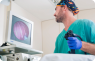 EndoscopyAn accurate diagnosis without conventional surgery.
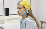 ElectrophysiologyFunctional exploration of the central nervous system.
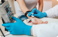 ElectromyographyClinical and neurophysiological evaluation of neuromuscular pathology.
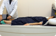 DensitometryDiagnostic technique for checking bone mineral density.
 UrodynamicsDiagnosis of urination disorders and incontinence.
- Medical check-ups

 GeneralThe most intelligent health check-up
 FullA comprehensive examination of your health
 Full PlusOur most exclusive check-up
 TravellersWhen travelling, your health is also part of your luggage
 SportA thorough review to boost your performance
 CardiologyGood news is knowing your heart is under control
 For companiesA tool that enhances employee satisfaction, productivity, and loyalty
- Diagnostic tests
- Our centre
- Teknon Healthcare Service Areas
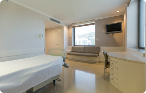 InpatientBright, functional and fully equipped rooms.
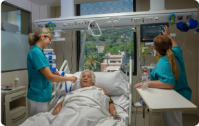 Semi-critical Care UnitEquipped with technology for diagnoses and treatments that require special care.
 Healthy Nutrition ProgrammeWe want to improve people’s health, which is why we promote healthy, conscious and sustainable nutrition at our hospitals.
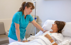 NursingOver 400 professionals.
 Emergency DepartmentUninterrupted operation, 24/7 at your service.
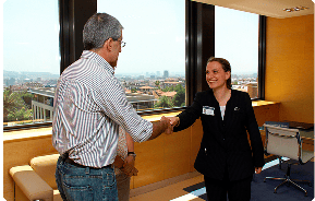 Exclusivity / Teknon ClubCommitted to superior and individualised service, we furnish a full range of services in conjunction with our medical and healthcare support.
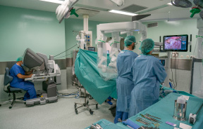 Surgical AreaA total of twenty (20) operating theatres, 12 of which are equipped for high-risk surgery.
 International ProgrammeA programme agent will provide you with comprehensive, personalised support
 Healthcare Ethics CommitteeGuidance for citizens and professionals in cases of moral conflicts.
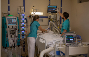 ICU-CCUMultipurpose unit incorporating treatment cubicles equipped with modern monitoring systems.
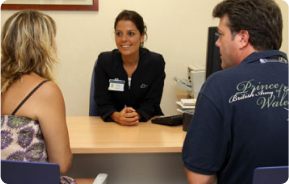 Patient servicesAvailable to all our patients and their companions.
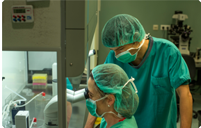 ResearchResearch is one of the cornerstones of Centro Médico Teknon.
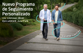 Personalised Follow-up ProgrammeWe accompany you throughout your medical journey. We organise your appointments and tests.
 Patient Quality and SafetyOur management models are based on the most stringent national and international standards.
- Teknon Healthcare Service Areas
- News
- News
 NewsKeep abreast of the events at Centro Médico Teknon. Visit our News section.
 AgendaHere we post upcoming events and discussions on relevant health topics. Visit our Agenda section to see what’s up.
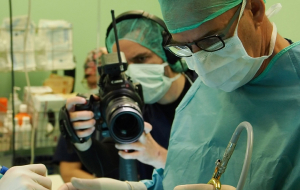 VideosThis section contains an extensive collection of videos related to our specialities.
 PodcastOur specialists discuss current medical topics, innovative treatments, health advice and patient experience.
 Health content
- News
- Blog

- Medical directory
- Specialities
- Specialised Units
- Teknon Cardiology Institute
- Obesity Unit
- Teknon Oncology Institute
- Bloodless Medicine and Surgery Unit
- Teknon Institute of Neurosciences
- Traffic Accident Care Unit
- Teknon Pulmonology Unit
- Maritime Medicine Unit
- Institute of Tissue Regenerative Therapy
- Teknon Pain Treatment Unit
- Teknon Tennis Clinic
- Systemic Inflammatory and Autoimmune Diseases Unit
- Assisted Reproduction Unit
- Sleep Unit
- Unidad de Síndromes de Sensibilización Central
- Specialised Units
- Diagnostics
- Diagnostic tests
- Diagnostic ImagingDiagnostic and interventional scans.
- Anatomical pathology laboratoryAllows to obtain a second opinion from renowned specialists.
- Clinical Analysis LaboratoryComprehensive service in the clinical area.
- EndoscopyAn accurate diagnosis without conventional surgery.
- ElectrophysiologyFunctional exploration of the central nervous system.
- ElectromyographyClinical and neurophysiological evaluation of neuromuscular pathology.
- DensitometryDiagnostic technique for checking bone mineral density.
- UrodynamicsDiagnosis of urination disorders and incontinence.
- Medical check-ups
- GeneralThe most intelligent health check-up
- FullA comprehensive examination of your health
- Full PlusOur most exclusive check-up
- TravellersWhen travelling, your health is also part of your luggage
- SportA thorough review to boost your performance
- CardiologyGood news is knowing your heart is under control
- For companiesA tool that enhances employee satisfaction, productivity, and loyalty
- Diagnostic tests
- Our centre
- Teknon Healthcare Service Areas
- InpatientBright, functional and fully equipped rooms.
- Semi-critical Care UnitEquipped with technology for diagnoses and treatments that require special care.
- Healthy Nutrition ProgrammeWe want to improve people’s health, which is why we promote healthy, conscious and sustainable nutrition at our hospitals.
- NursingOver 400 professionals.
- Emergency DepartmentUninterrupted operation, 24/7 at your service.
- Exclusivity / Teknon ClubCommitted to superior and individualised service, we furnish a full range of services in conjunction with our medical and healthcare support.
- Surgical AreaA total of twenty (20) operating theatres, 12 of which are equipped for high-risk surgery.
- International ProgrammeA programme agent will provide you with comprehensive, personalised support
- Healthcare Ethics CommitteeGuidance for citizens and professionals in cases of moral conflicts.
- ICU-CCUMultipurpose unit incorporating treatment cubicles equipped with modern monitoring systems.
- Patient servicesAvailable to all our patients and their companions.
- ResearchResearch is one of the cornerstones of Centro Médico Teknon.
- Personalised Follow-up ProgrammeWe accompany you throughout your medical journey. We organise your appointments and tests.
- Patient Quality and SafetyOur management models are based on the most stringent national and international standards.
- Teknon Healthcare Service Areas
- News
- News
- NewsKeep abreast of the events at Centro Médico Teknon. Visit our News section.
- AgendaHere we post upcoming events and discussions on relevant health topics. Visit our Agenda section to see what’s up.
- VideosThis section contains an extensive collection of videos related to our specialities.
- PodcastOur specialists discuss current medical topics, innovative treatments, health advice and patient experience.
- Health content
- News
- Blog


- Specialities
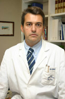
 Bardají Bofill Manel
- Servicios
- Abdomen
- Small intestine
- Gastrointestinal bleeding
- Bleeding from the upper digestive tract
 Centro Médico Teknonen/health-centers/centro-medico-teknon

Cause
In most cases it is due to peptic ulcer, gastritis, esophageal varices, esophagitis or gastric neoplasms. Peptic ulcer explains 50 to 67% of cases. The ratio of duodenal and gastric ulcers is 4:1. Hemorrhage is the initial symptom in 15% of cases. Surgical intervention is required only in 10 to 20% of patients. There are marginal ulcers after some gastric resections. Mortality reaches 10 to 20% and depends on age. Acute mucosal lesions are erosions of the mucosa, other than ulcers. Stress ulcers are seen in shock, septicemia, burn (Curling's ulcers), trauma, craniotomy (Cushing's ulcers), and major operations, and can be caused by decreased splanchnic flow and ischemic injury. Substances such as steroids, alcohol and aspirin produce erosive hemorrhagic gastritis. Esophageal varices account for 10% of all upper gastrointestinal tract hemorrhages and 95% massive hematemesis in children. They are a consequence of cirrhosis and portal hypertension (in the case of children, extrahepatic) and are more intense than alcoholic gastritis. Other lesions are hiatal hernia, gastric carcinoma, leiomyomas, vascular lesions, Mallory-Weiss tears, hematobilia and duodenal diverticula.
Diagnosis
- History and physical examination. The history should include medical conditions, especially ulceropathy, as well as the intensity and frequency of vomiting (Mallory-Weiss tear). Drug ingestion, bleeding tendencies, previous stomach operations, reflux symptoms and recent trauma should be investigated. The examination should detect the stigmas of causative diseases (cirrhosis and vascular lesions).
- Diagnostic methods. Hematocrit will usually not demonstrate the extent of the loss in acute cases. The SMA7 profile (increase in urea nitrogen by absorption of blood products in gastrointestinal viscera and by dehydration) includes measurement of ammonia in blood. Coagulation and platelet count studies must be done. Nasogastric aspiration of bilious but non-bloody fluid rules out proximal hemorrhage to the treitz ligament; while a clear, but not bilious, liquid does not rule it out.
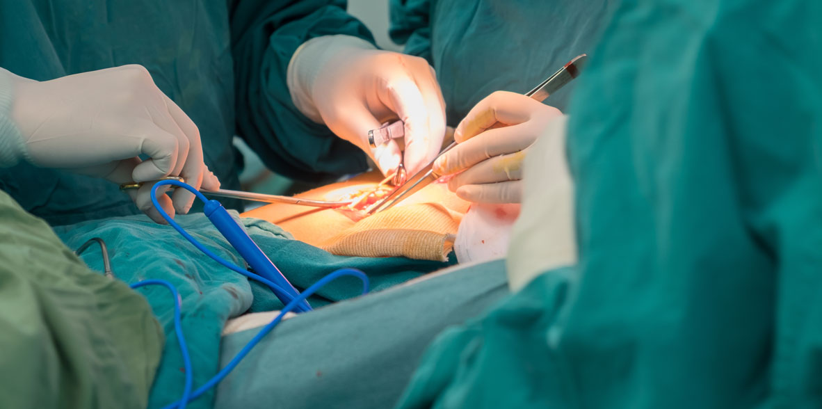
Endoscopy is an indispensable technique in the hemorrhage of the upper gastrointestinal tract. The study with red blood cells labeled with TC is useful in case of intermittent or low-intensity bleeding (0.1 ml/min). Angiography of the celiac trunk and superior mesenteric artery is useful in bleeding exceeding 1 to 2 ml/minute and 90% of the time is accurate. Selective embolization can be used at that time. Arterial vasopressin through these catheters is no more effective than intravenous administration. Gastroduodenal series may be done if no data are detected by endoscope or arteriography. Finally, diagnostic laparotomy should be considered, especially with massive bleeding. The long gastrostomy is used to inspect the duodenum, stomach, and hiatus.