

- Medical directory
- Specialities
- Specialised Units
 Teknon Cardiology Institute
 Obesity Unit
 Teknon Oncology Institute
 Bloodless Medicine and Surgery Unit
 Teknon Institute of Neurosciences
 Traffic Accident Care Unit
 Teknon Pulmonology Unit
 Maritime Medicine Unit
 Institute of Tissue Regenerative Therapy
 Teknon Pain Treatment Unit
 Teknon Tennis Clinic
 Systemic Inflammatory and Autoimmune Diseases Unit
 Assisted Reproduction Unit
 Sleep Unit
 Unidad de Síndromes de Sensibilización Central
- Specialised Units
- Diagnostics
- Diagnostic tests
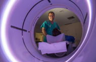 Diagnostic ImagingDiagnostic and interventional scans.
 Anatomical pathology laboratoryAllows to obtain a second opinion from renowned specialists.
 Clinical Analysis LaboratoryComprehensive service in the clinical area.
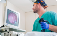 EndoscopyAn accurate diagnosis without conventional surgery.
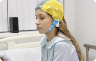 ElectrophysiologyFunctional exploration of the central nervous system.
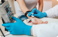 ElectromyographyClinical and neurophysiological evaluation of neuromuscular pathology.
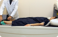 DensitometryDiagnostic technique for checking bone mineral density.
 UrodynamicsDiagnosis of urination disorders and incontinence.
- Medical check-ups

 GeneralThe most intelligent health check-up
 FullA comprehensive examination of your health
 Full PlusOur most exclusive check-up
 TravellersWhen travelling, your health is also part of your luggage
 SportA thorough review to boost your performance
 CardiologyGood news is knowing your heart is under control
 For companiesA tool that enhances employee satisfaction, productivity, and loyalty
- Diagnostic tests
- Our centre
- Teknon Healthcare Service Areas
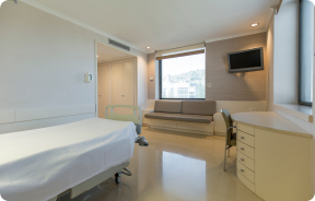 InpatientBright, functional and fully equipped rooms.
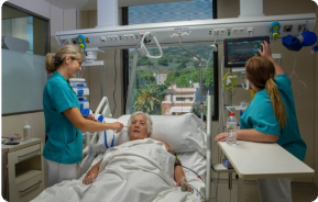 Semi-critical Care UnitEquipped with technology for diagnoses and treatments that require special care.
 Healthy Nutrition ProgrammeWe want to improve people’s health, which is why we promote healthy, conscious and sustainable nutrition at our hospitals.
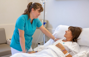 NursingOver 400 professionals.
 Emergency DepartmentUninterrupted operation, 24/7 at your service.
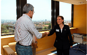 Exclusivity / Teknon ClubCommitted to superior and individualised service, we furnish a full range of services in conjunction with our medical and healthcare support.
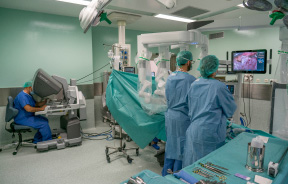 Surgical AreaA total of twenty (20) operating theatres, 12 of which are equipped for high-risk surgery.
 International ProgrammeA programme agent will provide you with comprehensive, personalised support
 Healthcare Ethics CommitteeGuidance for citizens and professionals in cases of moral conflicts.
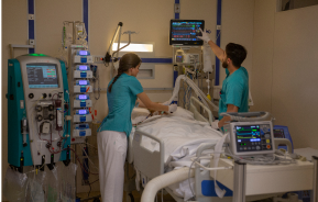 ICU-CCUMultipurpose unit incorporating treatment cubicles equipped with modern monitoring systems.
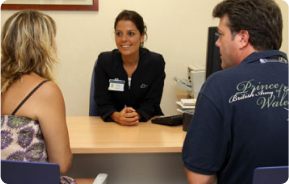 Patient servicesAvailable to all our patients and their companions.
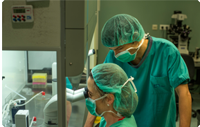 ResearchResearch is one of the cornerstones of Centro Médico Teknon.
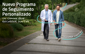 Personalised Follow-up ProgrammeWe accompany you throughout your medical journey. We organise your appointments and tests.
 Patient Quality and SafetyOur management models are based on the most stringent national and international standards.
- Teknon Healthcare Service Areas
- News
- News
 NewsKeep abreast of the events at Centro Médico Teknon. Visit our News section.
 AgendaHere we post upcoming events and discussions on relevant health topics. Visit our Agenda section to see what’s up.
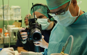 VideosThis section contains an extensive collection of videos related to our specialities.
 PodcastOur specialists discuss current medical topics, innovative treatments, health advice and patient experience.
 Health content
- News
- Blog

- Medical directory
- Specialities
- Specialised Units
- Teknon Cardiology Institute
- Obesity Unit
- Teknon Oncology Institute
- Bloodless Medicine and Surgery Unit
- Teknon Institute of Neurosciences
- Traffic Accident Care Unit
- Teknon Pulmonology Unit
- Maritime Medicine Unit
- Institute of Tissue Regenerative Therapy
- Teknon Pain Treatment Unit
- Teknon Tennis Clinic
- Systemic Inflammatory and Autoimmune Diseases Unit
- Assisted Reproduction Unit
- Sleep Unit
- Unidad de Síndromes de Sensibilización Central
- Specialised Units
- Diagnostics
- Diagnostic tests
- Diagnostic ImagingDiagnostic and interventional scans.
- Anatomical pathology laboratoryAllows to obtain a second opinion from renowned specialists.
- Clinical Analysis LaboratoryComprehensive service in the clinical area.
- EndoscopyAn accurate diagnosis without conventional surgery.
- ElectrophysiologyFunctional exploration of the central nervous system.
- ElectromyographyClinical and neurophysiological evaluation of neuromuscular pathology.
- DensitometryDiagnostic technique for checking bone mineral density.
- UrodynamicsDiagnosis of urination disorders and incontinence.
- Medical check-ups
- GeneralThe most intelligent health check-up
- FullA comprehensive examination of your health
- Full PlusOur most exclusive check-up
- TravellersWhen travelling, your health is also part of your luggage
- SportA thorough review to boost your performance
- CardiologyGood news is knowing your heart is under control
- For companiesA tool that enhances employee satisfaction, productivity, and loyalty
- Diagnostic tests
- Our centre
- Teknon Healthcare Service Areas
- InpatientBright, functional and fully equipped rooms.
- Semi-critical Care UnitEquipped with technology for diagnoses and treatments that require special care.
- Healthy Nutrition ProgrammeWe want to improve people’s health, which is why we promote healthy, conscious and sustainable nutrition at our hospitals.
- NursingOver 400 professionals.
- Emergency DepartmentUninterrupted operation, 24/7 at your service.
- Exclusivity / Teknon ClubCommitted to superior and individualised service, we furnish a full range of services in conjunction with our medical and healthcare support.
- Surgical AreaA total of twenty (20) operating theatres, 12 of which are equipped for high-risk surgery.
- International ProgrammeA programme agent will provide you with comprehensive, personalised support
- Healthcare Ethics CommitteeGuidance for citizens and professionals in cases of moral conflicts.
- ICU-CCUMultipurpose unit incorporating treatment cubicles equipped with modern monitoring systems.
- Patient servicesAvailable to all our patients and their companions.
- ResearchResearch is one of the cornerstones of Centro Médico Teknon.
- Personalised Follow-up ProgrammeWe accompany you throughout your medical journey. We organise your appointments and tests.
- Patient Quality and SafetyOur management models are based on the most stringent national and international standards.
- Teknon Healthcare Service Areas
- News
- News
- NewsKeep abreast of the events at Centro Médico Teknon. Visit our News section.
- AgendaHere we post upcoming events and discussions on relevant health topics. Visit our Agenda section to see what’s up.
- VideosThis section contains an extensive collection of videos related to our specialities.
- PodcastOur specialists discuss current medical topics, innovative treatments, health advice and patient experience.
- Health content
- News
- Blog


- Specialities
 Castro Domínguez Francisco
- Datos de interés
- Frequent questions
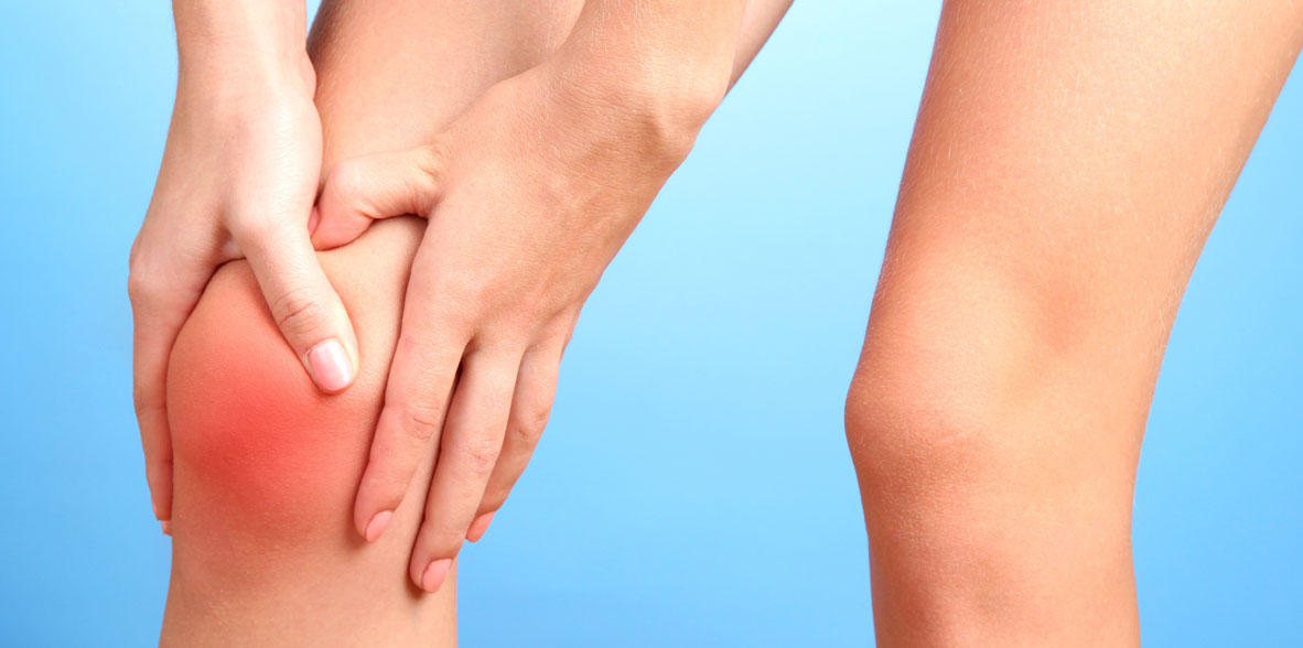
- Lumbar spinal stenosis: What are the presentation symptoms, how is it diagnosed and how is it treated?
 Centro Médico Teknonen/health-centers/centro-medico-teknon
- Centro Médico Teknonen/health-centers/centro-medico-teknonHospital Universitari General de Catalunyaen/health-centers/hospital-universitari-general-catalunya
 Centro Médico Teknonen/health-centers/centro-medico-teknonHospital Universitari Sagrat Coren/health-centers/hospital-universitari-sagrat-cor

Lumbar spinal stenosis is a medical condition characterized by the narrowing of the spinal canal in the lumbar region of the spine. This narrowing can lead to compression of the spinal cord and nerve roots, resulting in various symptoms.
Presentation Symptoms:
Neurogenic Claudication: This is a hallmark symptom of lumbar stenosis. Patients typically experience pain, cramping, or weakness in the lower back, buttocks, and legs. The discomfort often worsens with walking or prolonged standing and is relieved with sitting or bending forward.
Radicular Pain: Lumbar stenosis can cause radiating pain along the path of the affected nerves. Patients may experience pain, tingling, or numbness that can extend into the thighs, calves, and feet.
Sensory Changes: Some individuals may notice sensory changes in the legs, such as a "pins and needles" sensation or a feeling of heaviness.
Muscle Weakness: Stenosis can lead to muscle weakness in the lower extremities, potentially causing difficulty with walking and balance.
Loss of Bladder or Bowel Control: Although rare, severe lumbar stenosis can compress the cauda equina, a bundle of spinal nerves in the lower back, resulting in loss of bladder or bowel control. This is a medical emergency and requires immediate attention.
Diagnosis:
Clinical Evaluation: A thorough medical history and physical examination are essential for diagnosing lumbar stenosis. The healthcare provider will assess symptoms, risk factors, and neurological function. Imaging Studies: X-rays: X-rays can reveal structural changes in the spine, such as bone spurs and narrowing of the spinal canal.
Magnetic Resonance Imaging (MRI): MRI is a highly effective imaging technique for visualizing soft tissues, nerves, and the spinal canal. It can identify the degree of stenosis and any nerve compression.
Computed Tomography (CT) Scan: CT scans may be used to provide detailed cross-sectional images of the spine and bony structures.
Electrodiagnostic Tests:
Electromyography (EMG): EMG measures muscle activity and can help determine if there is nerve damage or compression. Nerve Conduction Studies: These tests assess the speed of electrical impulses through nerves, helping to identify areas of dysfunction.
Treatment:
Conservative Management
Physical Therapy: Physical therapy can help improve strength, flexibility, and posture, relieving symptoms and enhancing mobility. Medications: Nonsteroidal anti-inflammatory drugs (NSAIDs), muscle relaxants, and neuropathic pain medications may be prescribed to manage pain and inflammation.
Epidural Steroid Injections: These injections deliver anti-inflammatory steroids directly into the spinal area to reduce inflammation and alleviate symptoms.
Surgical Interventions:
Laminectomy: This surgical procedure involves the removal of bone and tissue to create more space in the spinal canal, relieving pressure on the nerves.
Laminotomy: Similar to laminectomy, but a smaller portion of bone is removed.
Spinal Fusion: Spinal fusion may be performed in conjunction with decompressive surgery to stabilize the spine and prevent further issues.
Minimally Invasive Surgery: In select cases, minimally invasive techniques, such as microdiscectomy or endoscopic decompression, may be used to minimize tissue damage and shorten recovery time.