


- Medical directory
- Specialities
- Unidades especializadas
 Instituto del Corazón
 Unidad de Obesidad
 Instituto Oncológico
 Unidad de Medicina y Cirugía sin sangre
 Instituto de Neurociencias
 Unidad de Atención al Lesionado de Tráfico
 Instituto de Neumología
 Unidad de Medicina Marítima
 Instituto de Terapia Regenerativa Tisular
 Unidad de Tratamiento del Dolor
 Clínica del Tenis
 Unidad de Enfermedades Inflamatorias y Autoinmunes
 Unidad de Reproducción
Asistida
Unidad de Reproducción
Asistida Unidad del Sueño
 Unidad de Síndromes de Sensibilización Central
- Unidades especializadas
- Diagnostics
- Diagnostic tests
 Diagnostic ImagingDiagnostic and interventional scans.
 Anatomical pathology laboratoryAllows to obtain a second opinion from renowned specialists.
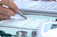 Clinical Analysis LaboratoryComprehensive service in the clinical area.
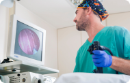 EndoscopyAn accurate diagnosis without conventional surgery.
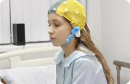 ElectrophysiologyFunctional exploration of the central nervous system.
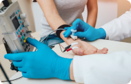 ElectromyographyClinical and neurophysiological evaluation of neuromuscular pathology.
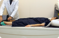 DensitometryDiagnostic technique for checking bone mineral density.
 UrodynamicsDiagnosis of urination disorders and incontinence.
- Chequeos médicos

 GeneralUn control inteligente de tu salud
 CompletoUn examen exhaustivo de tu salud
 Completo PlusNuestro chequeo más exclusivo
 ViajerosSi vas a emprender un viaje, tu salud es parte del equipaje
 DeportivoUna revisión a fondo para potenciar tu rendimiento
 CardiológicoUna buena noticia es saber que tu corazón está bajo control
 Para empresasUna herramienta que potencia la satisfacción, productividad y fidelización del empleado
- Diagnostic tests
- Our centre
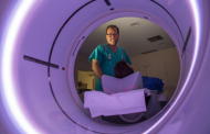
- Teknon Healthcare Service Areas
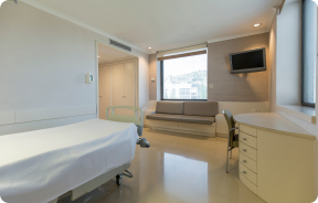 InpatientBright, functional and fully equipped rooms.
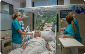 Semi-critical Care UnitEquipped with technology for diagnoses and treatments that require special care.
 Healthy Nutrition ProgrammeWe want to improve people’s health, which is why we promote healthy, conscious and sustainable nutrition at our hospitals.
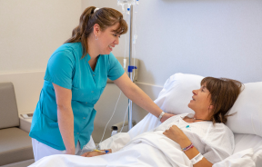 NursingOver 400 professionals.
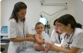 Emergency DepartmentUninterrupted operation, 24/7 at your service.
 Exclusivity / Teknon ClubCommitted to superior and individualised service, we furnish a full range of services in conjunction with our medical and healthcare support.
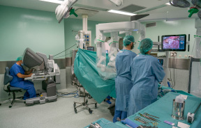 Surgical AreaA total of twenty (20) operating theatres, 12 of which are equipped for high-risk surgery.
 International ProgrammeA programme agent will provide you with comprehensive, personalised support
 Healthcare Ethics CommitteeGuidance for citizens and professionals in cases of moral conflicts.
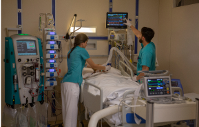 ICU-CCUMultipurpose unit incorporating treatment cubicles equipped with modern monitoring systems.
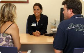 Patient servicesAvailable to all our patients and their companions.
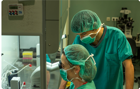 ResearchResearch is one of the cornerstones of Centro Médico Teknon.
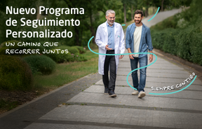 Programa de Seguimiento PersonalizadoTe acompañamos durante tu proceso médico. Organizamos y agendamos tus citas y pruebas.
 Calidad y Seguridad del PacienteAdoptamos modelos de gestión basados en los estándares más exigentes nacionales e internacionales.
- Teknon Healthcare Service Areas
- News
- Actualidad
 NoticiasConoce qué está pasando en Centro Médico Teknon. Consulta nuestra sección de noticias.
 AgendaPuedes encontrar todos los eventos que hemos organizado sobre salud y aquellos temas de actualidad que te pueden interesar. Accede a nuestra agenda de actividades.
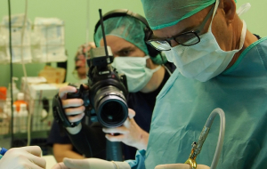 VídeosEn esta sección encontrarás una amplia colección de videos relacionados con nuestras especialidades.
 PodcastTemas médicos de actualidad, tratamientos innovadores, consejos de salud y experiencias de pacientes abordados por nuestros especialistas.
 Contenidos de salud
- Actualidad
- Blog

- Medical directory
- Specialities
- Unidades especializadas
- Instituto del Corazón
- Unidad de Obesidad
- Instituto Oncológico
- Unidad de Medicina y Cirugía sin sangre
- Instituto de Neurociencias
- Unidad de Atención al Lesionado de Tráfico
- Instituto de Neumología
- Unidad de Medicina Marítima
- Instituto de Terapia Regenerativa Tisular
- Unidad de Tratamiento del Dolor
- Clínica del Tenis
- Unidad de Enfermedades Inflamatorias y Autoinmunes
- Unidad de Reproducción
Asistida
- Unidad del Sueño
- Unidad de Síndromes de Sensibilización Central
- Unidades especializadas
- Diagnostics
- Diagnostic tests
- Diagnostic ImagingDiagnostic and interventional scans.
- Anatomical pathology laboratoryAllows to obtain a second opinion from renowned specialists.
- Clinical Analysis LaboratoryComprehensive service in the clinical area.
- EndoscopyAn accurate diagnosis without conventional surgery.
- ElectrophysiologyFunctional exploration of the central nervous system.
- ElectromyographyClinical and neurophysiological evaluation of neuromuscular pathology.
- DensitometryDiagnostic technique for checking bone mineral density.
- UrodynamicsDiagnosis of urination disorders and incontinence.
- Chequeos médicos
- GeneralUn control inteligente de tu salud
- CompletoUn examen exhaustivo de tu salud
- Completo PlusNuestro chequeo más exclusivo
- ViajerosSi vas a emprender un viaje, tu salud es parte del equipaje
- DeportivoUna revisión a fondo para potenciar tu rendimiento
- CardiológicoUna buena noticia es saber que tu corazón está bajo control
- Para empresasUna herramienta que potencia la satisfacción, productividad y fidelización del empleado
- Diagnostic tests
- Our centre
- Teknon Healthcare Service Areas
- InpatientBright, functional and fully equipped rooms.
- Semi-critical Care UnitEquipped with technology for diagnoses and treatments that require special care.
- Healthy Nutrition ProgrammeWe want to improve people’s health, which is why we promote healthy, conscious and sustainable nutrition at our hospitals.
- NursingOver 400 professionals.
- Emergency DepartmentUninterrupted operation, 24/7 at your service.
- Exclusivity / Teknon ClubCommitted to superior and individualised service, we furnish a full range of services in conjunction with our medical and healthcare support.
- Surgical AreaA total of twenty (20) operating theatres, 12 of which are equipped for high-risk surgery.
- International ProgrammeA programme agent will provide you with comprehensive, personalised support
- Healthcare Ethics CommitteeGuidance for citizens and professionals in cases of moral conflicts.
- ICU-CCUMultipurpose unit incorporating treatment cubicles equipped with modern monitoring systems.
- Patient servicesAvailable to all our patients and their companions.
- ResearchResearch is one of the cornerstones of Centro Médico Teknon.
- Programa de Seguimiento PersonalizadoTe acompañamos durante tu proceso médico. Organizamos y agendamos tus citas y pruebas.
- Calidad y Seguridad del PacienteAdoptamos modelos de gestión basados en los estándares más exigentes nacionales e internacionales.
- Teknon Healthcare Service Areas
- News
- Actualidad
- NoticiasConoce qué está pasando en Centro Médico Teknon. Consulta nuestra sección de noticias.
- AgendaPuedes encontrar todos los eventos que hemos organizado sobre salud y aquellos temas de actualidad que te pueden interesar. Accede a nuestra agenda de actividades.
- VídeosEn esta sección encontrarás una amplia colección de videos relacionados con nuestras especialidades.
- PodcastTemas médicos de actualidad, tratamientos innovadores, consejos de salud y experiencias de pacientes abordados por nuestros especialistas.
- Contenidos de salud
- Actualidad
- Blog

- Especialidades
 Fernández Agrafojo Dora
- Our services
- Retina (AMD)
 Ophthalmology
 OftalmologíaCentro Médico Teknon
 OphthalmologyCentro Médico Teknon
 OphthalmologyCentro Médico Teknon
 OftalmologíaCentro Médico Teknon
 OftalmologíaCentro Médico Teknon

The retina, innermost layer of the eye globe, is a transparent tissue composed of many photosensitive cells which receive light stimuli and transmit them through nerve terminals to the brain. There are 2 types of photoreceptor cells: cones and rods.
Cones function better in daylight and are specialized for color vision. Rods are more numerous and function in evening light or darkness. Cones are more plentiful in the center of the retina, also called the macula or fovea, and rods are found in the periphery. When cones and rods are stimulated by light, they generate impulses which are transmitted through their own nerve fibers, which in turn come together to form the optic nerve.
The retina is fed by retinal artery vessels and by capillaries of the choroid, the outermost vascularized layer of the retina.
- The vitreous
The vitreous is a transparent, gelatinous substance which occupies the inner part of the eye, providing support to the retina. It has no nutritive vessels, since it would lose its transparency. Over the years, the vitreous may lose its consistency and separate from the retina, causing deposits which are perceived as threads or spots that move with the eyes; these are more visible in bright light or when looking at light-colored walls or objects. These deposits are referred to as myodesopsia, "floaters", or "mouches volantes". Usually they do not indicate any kind of disease, nor do they represent any risk to one's vision; however, if they are perceived, one must rule out the presence of any coexisting pathology in the retina or vitreous.
- What are the main problems of the retina?
The retina can be affected by:
- Many general diseases, such as diabetes or ocular hypertension (high eye pressure)
- Inflammations or infections
- Vascular irregularities such as thrombosis or embolisms
- detachments which produce loss of vision in one part of the field of vision, or in its entirety
- Degeneration, its most frequent and important instance being age-related macular degeneration
- How to diagnose these diseases?
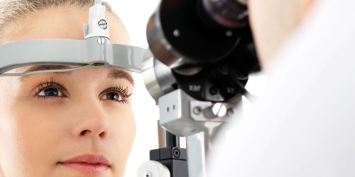
Diagnosis of all these diseases is important in order to provide early treatment. For this purpose we have many techniques and instruments at our disposal, most notably: retinoscopy (observation of the retina and vitreous with or without dilating the pupil), fluorescein angiography, computerized campimetry, and for some time now, optical coherence tomography – a very high-precision, latest-generation instrument for studying the retina.
Irregularities in the layers of the retina which nourish and provide oxygen to the pigment epithelium are the cause of age-related macular degeneration (AMD). This can be either dry or wet (exudative) AMD, the latter being characterized by the appearance of blood vessels, which can give rise to hemorrhaging and accumulation of liquid between layers of the retina.
With AMD, the patient will report irregularity in the central vision of the affected eye, image distortion or deformity.In such cases, the Amsler test offers a simple method for monitoring symptoms.
Recent years have brought to light the so-called antiangiogenic substances. When injected into the inner eye, they often make it possible to control loss of vision or even to improve it, in some cases of exudative (wet) age-related macular degeneration.
With the help of the latest technology in ophthalmological exploration, we are able to detect retinopathies in the earliest stages of macular degeneration, when the patient may not report any kind of symptoms. This is the case with Optical Coherence Tomography (OCT).
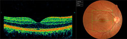
This technique examines retinal layers through images, visualizing a cross-section of the desired area of the retina. The slightest irregularity among the layers (as in cases of AMD) is projected, measured, compared and monitored, thanks to the different visual perspectives offered by the software. Even 3-D imaging of the retina can be obtained.
A database of population studies is incorporated, giving us automatic comparisons of the thickness of retinal ganglion fibers which lead to the optic nerve, and in this way assess their normality. This is especially useful in the case of patients with glaucoma, both as a support to diagnosis as well as in monitoring its development over time.