


- Cuadro médico
- Especialidades
- Unidades especializadas
 Instituto del Corazón
 Unidad de Obesidad
 Instituto Oncológico
 Unidad de Medicina y Cirugía sin sangre
 Instituto de Neurociencias
 Unidad de Atención al Lesionado de Tráfico
 Instituto de Neumología
 Unidad de Medicina Marítima
 Instituto de Terapia Regenerativa Tisular
 Unidad de Tratamiento del Dolor
 Clínica del Tenis
 Unidad de Enfermedades Inflamatorias y Autoinmunes
 Unidad de Reproducción
Asistida
Unidad de Reproducción
Asistida Unidad del Sueño
 Unidad de Síndromes de Sensibilización Central
- Unidades especializadas
- Área diagnóstica
- Diagnostic tests
 Diagnostic ImagingDiagnostic and interventional scans.
 Anatomical pathology laboratoryAllows to obtain a second opinion from renowned specialists.
 Clinical Analysis LaboratoryComprehensive service in the clinical area.
 EndoscopyAn accurate diagnosis without conventional surgery.
 ElectrophysiologyFunctional exploration of the central nervous system.
 ElectromyographyClinical and neurophysiological evaluation of neuromuscular pathology.
 DensitometryDiagnostic technique for checking bone mineral density.
 UrodynamicsDiagnosis of urination disorders and incontinence.
- Chequeos médicos

 GeneralUn control inteligente de tu salud
 CompletoUn examen exhaustivo de tu salud
 Completo PlusNuestro chequeo más exclusivo
 ViajerosSi vas a emprender un viaje, tu salud es parte del equipaje
 DeportivoUna revisión a fondo para potenciar tu rendimiento
 CardiológicoUna buena noticia es saber que tu corazón está bajo control
 Para empresasUna herramienta que potencia la satisfacción, productividad y fidelización del empleado
- Diagnostic tests
- Nuestro Centro
- Entorno asistencial Teknon
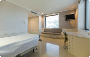 HospitalizaciónHabitaciones luminosas, funcionales y completamente equipadas.
 Unidad de SemicríticosDotada de tecnología para diagnósticos y tratamientos que requieren cuidado especial.
 Programa de Alimentación SaludableQueremos mejorar la salud de las personas, por ello promovemos una alimentación saludable, consciente y sostenible en nuestros hospitales.
 EnfermeríaEquipo de más de 400 profesionales.
 Servicio de Urgencias24 horas al día a tu servicio sin interrupciones.
 Exclusividad / Club TeknonBasado en la alta calidad y el servicio personalizado, ofrecemos un conjunto de servicios que complementan los cuidados médico-asistenciales
 Área QuirúrgicaCuenta con 20 quirófanos, 12 de ellos preparados para cirugía de alto riesgo.
 Programa internacionalUn asistente de este programa le ofrecerá atención integral y personalizada
 Comité de Ética AsistencialAyuda a ciudadanos y profesionales a orientar su actuación en casos de conflictos morales.
 UCI-UCUnidad polivalente con boxes equipados con modernos sistemas de monitorización.
 Atención al pacienteA disposición de todos los pacientes y acompañantes del centro.
 InvestigaciónLa investigación constituye uno de los pilares básicos de Centro Médico Teknon.
 Programa de Seguimiento PersonalizadoTe acompañamos durante tu proceso médico. Organizamos y agendamos tus citas y pruebas.
 Calidad y Seguridad del PacienteAdoptamos modelos de gestión basados en los estándares más exigentes nacionales e internacionales.
- Entorno asistencial Teknon
- Actualidad
- Actualidad
 NoticiasConoce qué está pasando en Centro Médico Teknon. Consulta nuestra sección de noticias.
 AgendaPuedes encontrar todos los eventos que hemos organizado sobre salud y aquellos temas de actualidad que te pueden interesar. Accede a nuestra agenda de actividades.
 VídeosEn esta sección encontrarás una amplia colección de videos relacionados con nuestras especialidades.
 PodcastTemas médicos de actualidad, tratamientos innovadores, consejos de salud y experiencias de pacientes abordados por nuestros especialistas.
 Contenidos de salud
- Actualidad
- Blog

- Cuadro médico
- Especialidades
- Unidades especializadas
- Instituto del Corazón
- Unidad de Obesidad
- Instituto Oncológico
- Unidad de Medicina y Cirugía sin sangre
- Instituto de Neurociencias
- Unidad de Atención al Lesionado de Tráfico
- Instituto de Neumología
- Unidad de Medicina Marítima
- Instituto de Terapia Regenerativa Tisular
- Unidad de Tratamiento del Dolor
- Clínica del Tenis
- Unidad de Enfermedades Inflamatorias y Autoinmunes
- Unidad de Reproducción
Asistida
- Unidad del Sueño
- Unidad de Síndromes de Sensibilización Central
- Unidades especializadas
- Área diagnóstica
- Diagnostic tests
- Diagnostic ImagingDiagnostic and interventional scans.
- Anatomical pathology laboratoryAllows to obtain a second opinion from renowned specialists.
- Clinical Analysis LaboratoryComprehensive service in the clinical area.
- EndoscopyAn accurate diagnosis without conventional surgery.
- ElectrophysiologyFunctional exploration of the central nervous system.
- ElectromyographyClinical and neurophysiological evaluation of neuromuscular pathology.
- DensitometryDiagnostic technique for checking bone mineral density.
- UrodynamicsDiagnosis of urination disorders and incontinence.
- Chequeos médicos
- GeneralUn control inteligente de tu salud
- CompletoUn examen exhaustivo de tu salud
- Completo PlusNuestro chequeo más exclusivo
- ViajerosSi vas a emprender un viaje, tu salud es parte del equipaje
- DeportivoUna revisión a fondo para potenciar tu rendimiento
- CardiológicoUna buena noticia es saber que tu corazón está bajo control
- Para empresasUna herramienta que potencia la satisfacción, productividad y fidelización del empleado
- Diagnostic tests
- Nuestro Centro
- Entorno asistencial Teknon
- HospitalizaciónHabitaciones luminosas, funcionales y completamente equipadas.
- Unidad de SemicríticosDotada de tecnología para diagnósticos y tratamientos que requieren cuidado especial.
- Programa de Alimentación SaludableQueremos mejorar la salud de las personas, por ello promovemos una alimentación saludable, consciente y sostenible en nuestros hospitales.
- EnfermeríaEquipo de más de 400 profesionales.
- Servicio de Urgencias24 horas al día a tu servicio sin interrupciones.
- Exclusividad / Club TeknonBasado en la alta calidad y el servicio personalizado, ofrecemos un conjunto de servicios que complementan los cuidados médico-asistenciales
- Área QuirúrgicaCuenta con 20 quirófanos, 12 de ellos preparados para cirugía de alto riesgo.
- Programa internacionalUn asistente de este programa le ofrecerá atención integral y personalizada
- Comité de Ética AsistencialAyuda a ciudadanos y profesionales a orientar su actuación en casos de conflictos morales.
- UCI-UCUnidad polivalente con boxes equipados con modernos sistemas de monitorización.
- Atención al pacienteA disposición de todos los pacientes y acompañantes del centro.
- InvestigaciónLa investigación constituye uno de los pilares básicos de Centro Médico Teknon.
- Programa de Seguimiento PersonalizadoTe acompañamos durante tu proceso médico. Organizamos y agendamos tus citas y pruebas.
- Calidad y Seguridad del PacienteAdoptamos modelos de gestión basados en los estándares más exigentes nacionales e internacionales.
- Entorno asistencial Teknon
- Actualidad
- Actualidad
- NoticiasConoce qué está pasando en Centro Médico Teknon. Consulta nuestra sección de noticias.
- AgendaPuedes encontrar todos los eventos que hemos organizado sobre salud y aquellos temas de actualidad que te pueden interesar. Accede a nuestra agenda de actividades.
- VídeosEn esta sección encontrarás una amplia colección de videos relacionados con nuestras especialidades.
- PodcastTemas médicos de actualidad, tratamientos innovadores, consejos de salud y experiencias de pacientes abordados por nuestros especialistas.
- Contenidos de salud
- Actualidad
- Blog

- Unidades especializadas
 Diagnostic tests
- Treatments and Specialities
- Diagnostic Imaging
- Multidetector Computed Tomography
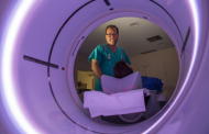
Multidetector Computed Tomography
Centro Médico Teknon uses state-of-the-art technology to render three-dimensional reconstruction of the organ under examination. Computed Tomography uses a rotational movement to send successive X-ray emissions to one or more detectors. This data is integrated into a single system which exports it in a ‘volume’ of images representing the scanned region. This provides detailed images with a density resolution far superior to that of conventional radiology.
- Neuroradiology
- Skull CT

Radiological test that provides high definition anatomical images of the skull (brain stem, cerebellum, cerebrum, cranial calotte, etc.) using CT (Computed Tomography) equipment. Indicated for: trauma, headache, memory disorders, sudden loss of strength in a limb or half of the body.
- Neck CT

Radiological test that provides high definition anatomical images of the neck using CT (Computed Tomography) equipment. Indicated for: thyroid study, control of treated tumours, study of lymph nodes, infections and abscesses.
- Laryngeal CT

Radiological test that provides high definition anatomical images of the larynx using CT (Computed Tomography) equipment. Indicated for: sudden or chronic aphonia, respiratory distress.
- Orbit CT

Radiological test that provides high definition anatomical images of the orbits using CT (Computed Tomography) equipment. Indicated for: double vision, infections, trauma, bulging eyes (hyperthyroidism), congenital malformations.
- Pituitary CT

Radiological test that provides high definition anatomical images of the cerebral pituitary gland using CT (Computed Tomography) equipment. Indicated for: suspected pituitary tumour, growth disorder.
- Facial mass CT


Radiological test that provides high definition anatomical images of the facial mass (face) using CT (Computed Tomography) equipment. Indicated for: tumours, plastic surgery.
- Ear CT

Radiological test that provides high definition anatomical images of the ear (internal and external auditory canal, eardrum, ossicles of the ear), using CT (Computed Tomography) equipment. Indicated for: hearing disorders, vertigo, dizziness, tinnitus (ringing).
- Dental CT

Radiological test that provides high definition anatomical images of the maxillary bone (teeth, dental nerve path) using CT (Computed Tomography) equipment. Indicated for: examination prior to dental extraction, examination prior to implants, tumours, abscess.
- Paranasal Sinuses CT

Radiological test that provides high definition anatomical images of the paranasal sinuses using CT (Computed Tomography) equipment. Indicated for: headache, mucus, facial infections.
- Temporal bone CT

Radiological test that provides high definition anatomical images of the temporal bone (inner, middle and outer ear) using CT (Computed Tomography) equipment. Indicated for: sudden or chronic hearing loss, vertigo, dizziness, congenital malformations.
- Angio – Supra-Aortic Trunk CT

Radiological test that provides high definition anatomical images of the carotid arteries of the neck using CT (Computed Tomography) equipment and the injection of an intravenous contrast agent. The images are then reconstructed in three dimensions (3D). Indicated for: acute cerebral vascular accident, transient vascular accident, carotid bruit.
- Cervical spine CT

Radiological test that provides high definition anatomical images of the cervical vertebrae using CT (Computed Tomography) equipment. Indicated for: cervical pain without/with irradiation to the arms, trauma.
- Thorax
- Chest CT

Diagnostic test that provides high definition anatomical images of the chest (lungs, heart, mediastinum, great vessels, rib cage, etc.) using CT (Computed Tomography) equipment. These images are then examined on a workstation that allows bidimensional reconstructions in different planes of space and also 3D reconstructions (volumetric). Some studies require the use of an iodinated contrast agent to improve image definition.
- Thoracic aorta CT angiography

Diagnostic test to examine the thoracic aorta (main artery of the thorax) using CT (Computed Tomography) equipment. This technique requires the use of an iodinated contrast agent, and provides high definition anatomical images. The use of MDCT (Multidetector Computed Tomography) shortens scanning time, reduces radiation dose and improves image quality. The multiple detectors used in certain studies enable imaging to be synchronised with the heartbeat, a technique used to study the aortic valve and aortic root (the first few centimetres), where the heartbeat often distorts images due to movement.
- Pulmonary Artery CT angiography (PTE Study, Pulmonary Thromboembolism)

Diagnostic test to examine the pulmonary arteries using CT (Computed Tomography) equipment to obtain two- and three-dimensional images. This requires the use of an iodinated contrast agent, which will provide improved anatomical definition. This test is mainly indicated in cases of suspected pulmonary thromboembolism (PTE) to rule out or confirm the presence of blood clots inside the arteries.
- High resolution Chest CT

Diagnostic test to examine the lung using CT (Computed Tomography) equipment to obtain two- and three-dimensional images, that allow a highly specific anatomical examination of the lung, being able to assess very small anatomical structures. This technique is very important among patients with suspected lung disease.
- Sternum CT

Utilising an X-ray system and detectors that rotate around a patient, this radiological scan generates images that are computer-reconstructed to facilitate a close examination of the sternum.
- Clavicle CT

Utilising an X-ray system and detectors that rotate around a patient, this radiological scan generates images that are computer-reconstructed to facilitate a close examination of the clavicles.
- Ribcage CT

Utilising an X-ray system and detectors that rotate around a patient, this radiological scan generates images that are computer-reconstructed to facilitate a close examination of the ribcage.
- Coronary CT angiography or Coronary CT

The Coronary CT angiography or non-invasive Coronarography is a diagnostic test to examine arteries of the heart, or coronary arteries, using state-of-the-art MDCT equipment and an iodinated contrast agent to obtain two- and three-dimensional images. Multidetector computed tomography (MDCT) entails high-speed imaging that is beneficial in assessing coronary arteries with high anatomical precision, particularly in evaluating narrowing or stenosis, calcifications, anatomical variants, etc., as its speed prevents the artefact caused by the constant movement of the heart (1,000 images can be obtained in less than 10 seconds). The information obtained requires processing at workstations equipped with specialised software capable of reconstructing the coronary arteries, thereby enabling an assessment to be made of the number, location and characteristics of the lesions. All this information is obtained non-invasively, involving a simple puncture of a peripheral vein (in the arm). To ensure the heart rate stays below 75 bpm, some patients will need preliminary treatment with beta blockers.
- CT-guided thoracic FNA (fine needle aspiration)

This test obtains a sample of tissue from thoracic lesions, such as lung masses, mediastinal masses, bone lesions, etc. This test is performed using local anaesthesia on the puncture area, which is administered with fine-gauge needles. The entire procedure is performed with guidance from images obtained by computed tomography (CT) at various stages of the puncture, using fluoroscopy-CT equipment. After the test, the patient remains in hospital for a few hours. Coagulation tests must be performed before the puncture.
- CT-guided thoracic biopsy

It consists of obtaining a tissue sample from a specific thoracic lesion, such as the lung, mediastinum, sternum, etc. It is sometimes performed under sedation with the help of an anaesthesia team. Needles are used to draw a cylinder sample from the lesion to be studied, which is then sent to the Pathology Department for histological analysis. The entire procedure is performed with guidance from images obtained by computed tomography (CT) at various stages of the biopsy, using fluoroscopy-CT equipment. After the test, the patient remains in hospital under observation. Coagulation tests must be performed before the puncture.
- Thoracic spine CT
Radiological test that involves capturing high-definition anatomical images of the thoracic vertebrae using a CT (computed tomography) scanner. Indicated for: acute/chronic back pain, trauma, spinal misalignment.
- Virtual bronchoscopy

Virtual bronchoscopy is a non-invasive technique for rendering three-dimensional and two-dimensional images of the trachea and bronchial tree through sequential imaging captured with a MDCT scanner. High-quality images enable virtual exploration inside the trachea and bronchi through processing on specialised workstations.
- Calcium score

Diagnostic test that measures the amount of calcium that may have accumulated in the atherosclerotic plaques of the coronary arteries using state-of-the-art MDCT equipment. It is a non-invasive test that does not require iodinated contrast. No prior preparation is necessary. Calcified plaques are detected using a specialised workstation that allows the exact amount of calcium to be quantified and given a score, which is known in medical practice as the calcium score.
- Abdomen and pelvis
- Abdomen CT

Diagnostic test that involves obtaining high-definition anatomical images of the abdomen (liver, gallbladder, bile duct, pancreas, spleen, stomach, intestines, kidneys, vascular structures, bladder, uterus and ovaries, etc.) using CT (computed tomography) equipment. These images are then studied at a workstation capable of producing two-dimensional reconstructions in different spatial planes, and also 3D (volumetric) reconstructions. Most studies require the use of iodinated contrast to improve image definition.
- Pelvis CT

Diagnostic test that involves obtaining high-definition anatomical two- and three-dimensional images of the pelvis (bone structures, vascular structures, bladder, uterus and ovaries, prostate and seminal vesicles, ureters, etc.) using CT (computed tomography) equipment. Most studies require the use of iodinated contrast.
- Abdominal and pelvic CT

Diagnostic test that consists of obtaining high-definition anatomical images (bone structures, vascular structures, liver, pancreas, gallbladder, kidneys, adrenal glands, spleen, small and large intestine, bladder, uterus and ovaries, prostate and seminal vesicles, ureters, etc.) using CT (computed tomography) equipment. Most studies require the use of iodinated contrast.
- Liver CT

Diagnostic test that involves obtaining high-definition anatomical two- and three-dimensional images of the liver using CT (computed tomography) equipment. The study must be performed before and after the use of iodinated contrast, carrying it out in different ‘hepatic phases’ to correctly assess all structures: hepatic parenchyma, intra- and extrahepatic bile ducts, gallbladder, hepatic vessels (hepatic artery, portal vein and suprahepatic veins) and adjacent structures (stomach, duodenum, inferior vena cava, pancreatic gland, etc.). This test is particularly useful in the study of liver damage, chronic liver disease, etc.
- Kidney CT

Diagnostic test that involves obtaining high-definition anatomical two- and three-dimensional images of the kidney and urinary system using CT (computed tomography) equipment. The study is performed before and after the use of iodinated contrast in different ‘renal phases’ for functional and anatomical assessment (renal parenchyma, ureters, urinary bladder, renal arteries and veins, etc.), as well as adjacent structures (inferior vena cava, abdominal aorta, liver, spleen, etc.). It is particularly recommended when kidney damage is suspected, in patients with blood in their urine or haematuria, etc.
- Urological CT

Diagnostic test that involves obtaining high-definition anatomical two- and three-dimensional images of the kidney and urinary system using CT (computed tomography) equipment. It is particularly recommended for patients suspected of having kidney stones, recurrent urinary tract infections, etc. The test is performed without using iodine contrast (only in certain cases will it be necessary to complete the test with iodine contrast).
- Pancreas CT

Diagnostic test that involves obtaining high-definition anatomical two- and three-dimensional images of the pancreas using CT (computed tomography) equipment. The study is performed before and after the use of iodinated contrast in different ‘pancreatic phases’ in order to assess all structures (pancreatic parenchyma, pancreatic duct or Wirsung duct, biliary-pancreatic junction, common bile duct, pancreatic arteries, splenic artery and vein, duodenum, etc.). It is particularly recommended when pancreatic injury is suspected, in patients with acute or chronic pancreatitis, etc.
- Abdominal aorta CT angiography


A non-invasive diagnostic test that involves studying the abdominal aorta by obtaining high-definition anatomical images using CT (computed tomography) equipment and iodinated contrast. With the aid of workstations specialised for arterial studies, the image quality supports 2D and 3D reconstructions. It is indicated in patients with vascular disease (atherosclerosis), aortic aneurysms, abdominal pain of possible vascular origin, pre-surgical studies of lesions adjacent to the abdominal aorta as a vascular ‘map’, etc. Information obtained non-invasively is indispensable for patients requiring percutaneous or surgical processing. In patients who only require tracking of vascular lesions, this technique is the non-invasive technique of choice, together with MRI angiography.
- Renal artery CT angiography


A non-invasive diagnostic test that involves studying the renal arteries by obtaining high-definition anatomical images using CT (computed tomography) equipment and iodinated contrast. With the aid of workstations specialised for arterial studies, the image quality supports 2D and 3D reconstructions. This test is recommended, for example, in patients suffering from refractory hypertension that does not respond to processing, in patients with kidney damage in order to obtain a pre-surgical ‘vascular’ map, etc.
- Aortoiliac CT angiography

A non-invasive diagnostic test that involves examining the iliac arteries and abdominal aorta, obtaining high-definition anatomical images using CT (computed tomography) equipment and iodinated contrast dye. With the aid of workstations specialised for arterial studies, the image quality supports 2D and 3D reconstructions. This test is particularly recommended as a pre-surgical study (vascular map) prior to percutaneous or surgical interventions on the abdominal aorta, as a complementary study in patients with lower limb ischaemia, etc.
- Virtual colonoscopy


Virtual colonoscopy is a non-invasive technique that allows three-dimensional and two-dimensional visualisation of the large intestine or colon by taking sequential images captured with a state-of-the-art MDCT scanner. The quality of the images allows virtual navigation through the rectum and colon thanks to processing on specialised workstations. Preparation for the test consists of following a low-fibre diet for three days before the test (to cleanse the colon and rectum) and ingesting an iodinated oral contrast agent the day before the test (to mark the stool so that it can be correctly distinguished from any colonic lesions). Unlike fibrocolonoscopy, no sedation or bowel preparation is required. The test is performed in the CT room, where air is blown through a small flexible tube to distend the colon.
- CT-guided abdominal FNA (fine needle aspiration)

It consists in obtaining a tissue sample from a specific lesion located in the abdominal cavity. This test is performed using local anaesthesia on the puncture area, which is administered with fine-gauge needles. The entire procedure is monitored using images obtained by computed tomography (CT) at various stages of the puncture, using fluoroscopy-CT equipment. After the test, the patient remains under observation in hospital for a few hours. Coagulation tests must be performed before the puncture.
- CT-guided abdominal biopsy

It consists in obtaining a tissue sample from a specific lesion located in the abdominal cavity. It is sometimes performed under sedation with the help of an anaesthesia team. Needles are used to draw a cylinder sample from the lesion to be studied, which is then sent to the Pathology Department for histological analysis. The entire procedure is monitored using images obtained by computed tomography (CT) at various stages of the biopsy, using CT fluoroscopy. After the test, the patient remains in hospital under observation. Coagulation tests must be performed before the puncture.
- CT-guided abdominal drainage (abscesses, collections)

It consists of placing a drainage catheter over a collection of fluid located in the abdominal cavity, with the intention of emptying as much of the collection as possible. The patient should keep the drain in place for a few days, usually until it is no longer productive. It is often performed under sedation with the help of an anaesthesia team. The entire procedure is monitored using images obtained by computed tomography (CT) at various stages of the test, using CT fluoroscopy. After the test, the patient remains hospitalised. Coagulation tests must be performed before the test.
- Osteoarticular
- Shoulder CT

Radiological examination based on an X-ray system and detectors that rotate around the patient, reconstructing the images by computer (multidetector computed tomography - MDCT) to study the bones, muscles and joints of the shoulder.
- Elbow CT

Radiological examination based on an X-ray system and detectors that rotate around the patient, reconstructing the images by computer (multidetector computed tomography - MDCT) to study the bones, muscles and joints of the elbow.
- Hand – wrist CT

Radiological examination based on an X-ray system and detectors that rotate around the patient, reconstructing the images by computer (multidetector computed tomography - MDCT) to study the bones, muscles and joints of the hands and wrists.
- Pelvic bone CT

Radiological examination based on an X-ray system and detectors that rotate around the patient, reconstructing the images by computer (multidetector computed tomography - MDCT) to study the bones, muscles and joints of the pelvis.
- Hip CT

Radiological examination based on an X-ray system and detectors that rotate around the patient, reconstructing the images by computer (multidetector computed tomography - MDCT) to study the bones, muscles and joints of the hips.
- Sacroiliac CT

Radiological examination based on an X-ray system and detectors that rotate around the patient, reconstructing the images by computer (multidetector computed tomography - MDCT) to study the sacroiliac joints and rule out inflammatory, traumatic or degenerative diseases.
- Knee CT

Radiological examination based on an X-ray system and detectors that rotate around the patient, reconstructing the images by computer (multidetector computed tomography - MDCT) to study the bones, muscles and joints of the knees.
- Ankle-toe CT

Radiological examination based on an X-ray system and detectors that rotate around the patient, reconstructing the images by computer (multidetector computed tomography - MDCT) to study the bones, muscles and joints of the ankle and foot.
- Lower leg rotational study using CT [patella, tibial tuberosity to trochlear groove (TT-TG) distance]

Radiological examination based on an X-ray system and detectors that rotate around the patient, reconstructing the images by computer (multidetector computed tomography - MDCT) to calculate a series of measurements at the hips, knees and ankles with a view to solving problems affecting the rotation and angulation of the lower limbs.
- Long bone CT

Radiological examination based on an X-ray system and detectors that rotate around the patient, reconstructing the images by computer (multidetector computed tomography - MDCT) to study the long bones (tibia, fibula, femur, humerus, radius and ulna).
- CT-guided bone biopsy

It consists of obtaining a tissue sample from a specific bone lesion. It is sometimes performed under sedation with the help of an anaesthesia team. Needles are used to draw a cylinder sample from the lesion to be studied, which is then sent to the Pathology Department for histological analysis. The entire procedure is monitored using images obtained by computed tomography (CT) at various stages of the biopsy, using CT fluoroscopy. After the test, the patient remains in hospital under observation. Coagulation tests must be performed before the puncture.
- Lower leg arterial CT angiography

Non-invasive diagnostic test consisting of a vascular study of the aorto-iliac sector and the arterial vessels of both lower extremities, obtaining high-definition anatomical images using state-of-the-art multidetector CT equipment and iodinated contrast. With the aid of workstations specialised for arterial studies, the image quality supports 2D and 3D reconstructions.
- Spinal column
- Cervical spine CT

Radiological test that provides high definition anatomical images of the cervical vertebrae using CT (Computed Tomography) equipment. Indicated for: neck pain with or without radiation to the arms, trauma, congenital malformations.
- Thoracic spine CT

Radiological test that involves capturing high-definition anatomical images of the thoracic vertebrae using a CT (computed tomography) scanner. Indicated for: back pain, study of spinal deviations, trauma.
- Lumbar spine CT

Radiological test that provides high definition anatomical images of the lumbar vertebrae using CT (Computed Tomography) equipment. Indicated for: lower back pain with or without radiation to the legs, difficulty walking, trauma.
- Sacrum-coccyx CT
- Paediatrics
- Skull CT

Radiological test that provides high definition anatomical images of the skull using CT (Computed Tomography) equipment. Indicated for: headache, tumour study, head trauma.
- Neck CT

Radiological test that provides high definition anatomical images of the neck using CT (Computed Tomography) equipment. Indicated for: infections, abscesses, lymph node examination.
- Chest CT

Diagnostic test that involves obtaining high-definition anatomical images of the chest (lungs, heart, mediastinum, large vessels, rib cage, etc.) using CT (computed tomography) equipment. These images are then studied at a workstation that allows two-dimensional reconstructions in different planes of space, as well as three-dimensional (3D: volumetric) reconstructions. Some studies require the use of an iodinated contrast agent to improve image definition.
- Abdomen CT

Diagnostic test that involves obtaining high-definition anatomical images of the abdomen (liver, gallbladder, bile duct, pancreas, spleen, stomach, intestines, kidneys, vascular structures, bladder, uterus and ovaries, etc.) using CT (computed tomography) equipment. These images are then studied at a workstation capable of producing two-dimensional reconstructions in different spatial planes, and also 3D (volumetric) reconstructions. Most studies require the use of iodinated contrast to improve image definition.
- Lower leg rotational study using CT
Radiological examination based on an X-ray system and detectors that rotate around the patient, reconstructing the images by computer to calculate a series of measurements at the hips, knees and ankles with a view to solving problems affecting the rotation and angulation of the lower limbs.
- Bone CT

Radiological test that provides high definition anatomical images of the bones using CT (Computed Tomography) equipment. Indicated for: trauma, study of focal bone lesions.
- CT under sedation

Radiological examination involving an X-ray system and detectors that rotate around the patient, reconstructing images by computer to study any region of the body. It is performed under sedation with the assistance of an anaesthesia team. It is extremely useful in paediatric patients, who can often be difficult to examine because of false images caused by movements (infants, young children, etc., when clinically indicated).
- Vascular studies
- Supra-aortic trunk CT angiography

Radiological test that provides high definition anatomical images of the carotid arteries of the neck using CT (Computed Tomography) equipment and the injection of an intravenous contrast agent. The images are then reconstructed in three dimensions (3D). Indicated for: acute cerebral vascular accident, transient vascular accident, carotid bruit.
- Thoracic aorta CT angiography

Diagnostic test to examine the thoracic aorta (main artery of the thorax) using CT (Computed Tomography) equipment. This technique provides high-definition anatomical images. In most cases, the use of iodinated contrast is necessary. The use of MDCT (Multidetector Computed Tomography) shortens scanning time, reduces radiation dose and improves image quality. The multiple detectors used in certain studies enable imaging to be synchronised with the heartbeat, a technique used to study the aortic valve and aortic root (the first few centimetres), where the heartbeat tends to distort images due to movement.
- Pulmonary Artery CT angiography (PTE Study, Pulmonary Thromboembolism)

Diagnostic test to examine the pulmonary arteries using CT (Computed Tomography) equipment to obtain two- and three-dimensional images. The use of iodinated contrast is essential in this study, as it will allow for better anatomical definition. This test is mainly indicated in cases of suspected pulmonary thromboembolism (PTE) to rule out or confirm the presence of blood clots inside the arteries.
- Coronary CT angiography

The Coronary CT angiography or non-invasive Coronarography is a diagnostic test to examine arteries of the heart, or coronary arteries, using state-of-the-art MDCT equipment (64 slices or rows of detectors) and an iodinated contrast agent to obtain two- and three-dimensional images. 64-slice multidetector computed tomography (MDCT) entails high-speed imaging that is beneficial in assessing coronary arteries with high anatomical precision, particularly in evaluating narrowing or stenosis, calcifications, anatomical variants, etc., as its speed prevents the artefact caused by the constant movement of the heart (1,000 images can be obtained in less than 10 seconds). The information obtained requires processing at workstations equipped with specialised software capable of reconstructing the coronary arteries, thereby enabling an assessment to be made of the number, location and characteristics of the lesions. All this information is obtained non-invasively, involving a simple puncture of a peripheral vein (in the arm). To ensure the heart rate stays below 75 bpm, some patients will need preliminary treatment with beta blockers.
- Abdominal aorta CT angiography

A non-invasive diagnostic test that involves studying the abdominal aorta by obtaining high-definition anatomical images using CT (computed tomography) equipment and iodinated contrast. With the aid of workstations specialised for arterial studies, the image quality supports 2D and 3D reconstructions. It is indicated in patients with vascular disease (atherosclerosis), aortic aneurysms, abdominal pain of possible vascular origin, pre-surgical studies of lesions adjacent to the abdominal aorta as a vascular ‘map’, etc. Information obtained non-invasively is indispensable for patients requiring percutaneous or surgical processing. In patients who only require tracking of vascular lesions, this technique is the non-invasive technique of choice, together with MRI angiography.
- Renal artery CT angiography

A non-invasive diagnostic test that involves studying the renal arteries by obtaining high-definition anatomical images using CT (computed tomography) equipment and iodinated contrast. With the aid of workstations specialised for arterial studies, the image quality supports 2D and 3D reconstructions. This test is recommended, for example, in patients suffering from refractory hypertension that does not respond to processing, in patients with kidney damage in order to obtain a pre-surgical ‘vascular’ map, etc.
- Aortoiliac CT angiography

A non-invasive diagnostic test that involves examining the iliac arteries and abdominal aorta, obtaining high-definition anatomical images using CT (computed tomography) equipment and iodinated contrast dye. With the aid of workstations specialised for arterial studies, the image quality supports 2D and 3D reconstructions. This test is particularly recommended as a pre-surgical study (vascular map) prior to percutaneous or surgical interventions on the abdominal aorta, as a complementary study in patients with lower limb ischaemia, etc.
- Lower leg arterial CT angiography

Non-invasive diagnostic test consisting of a vascular study of the aorto-iliac sector and the arterial vessels of both lower extremities, obtaining high-definition anatomical images using state-of-the-art multidetector CT equipment and iodinated contrast. With the aid of workstations specialised for arterial studies, the image quality supports 2D and 3D reconstructions.
- CT-guided interventional procedures
- CT-guided thoracic FNA (fine needle aspiration)

This test obtains a sample of tissue from thoracic lesions, such as lung masses, mediastinal masses, bone lesions, etc. This test is performed using local anaesthesia on the puncture area, which is administered with fine-gauge needles. The entire procedure is monitored using images obtained by computed tomography (CT) at various stages of the puncture, using CT fluoroscopy. After the test, the patient remains in hospital for a few hours. Coagulation tests must be performed before the puncture.
- CT-guided thoracic biopsy

It consists of obtaining a tissue sample from a specific thoracic lesion, such as the lung, mediastinum, sternum, etc. It is sometimes performed under sedation with the help of an anaesthesia team. Needles are used to draw a cylinder sample from the lesion to be studied, which is then sent to the Pathology Department for histological analysis. The entire procedure is monitored using images obtained by computed tomography (CT) at various stages of the biopsy, using CT fluoroscopy. After the test, the patient remains in hospital under observation. Coagulation tests must be performed before the puncture.
- CT-guided abdominal FNA (fine needle aspiration)

It consists in obtaining a tissue sample from a specific lesion located in the abdominal cavity. This test is performed using local anaesthesia on the puncture area, which is administered with fine-gauge needles. The entire procedure is monitored using images obtained by computed tomography (CT) at various stages of the puncture, using CT fluoroscopy. After the test, the patient remains under observation in hospital for a few hours. Coagulation tests must be performed before the test.
- CT-guided abdominal biopsy

It consists in obtaining a tissue sample from a specific lesion located in the abdominal cavity. It is sometimes performed under sedation with the help of an anaesthesia team. Needles are used to draw a cylinder sample from the lesion to be studied, which is then sent to the Pathology Department for histological analysis. The entire procedure is monitored using images obtained by computed tomography (CT) at various stages of the biopsy, using CT fluoroscopy. After the test, the patient remains in hospital under observation. Coagulation tests must be performed before the puncture.
- CT-guided abdominal drainage

It consists of placing a drainage catheter over a collection of fluid located in the abdominal cavity, with the intention of emptying as much of the collection as possible. The patient should keep the drain in place for a few days, usually until it is no longer productive. It is often performed under sedation with the help of an anaesthesia team. The entire procedure is monitored using images obtained by computed tomography (CT) at various stages of the test, using CT fluoroscopy. After the test, the patient remains hospitalised. Coagulation tests must be performed before the test.
- CT-guided bone biopsy

Invasive examination used to extract bone tissue samples, mainly from tumours. The CT scan is used to select the puncture site and guide the biopsy needles to the tumour. Local anaesthesia is applied and sedation is occasionally used. The duration of the procedure will depend on its technical difficulty.
- Gold seeding for radiotherapy

An invasive procedure that uses specific needles and equipment to insert a gold marker (measuring just a few millimetres) next to tumour lesions located in internal organs. These particles will subsequently facilitate the application of radiotherapy to the lesions, making it more effective and less harmful to healthy tissue. The procedure is guided by CT fluoroscopy. Local anaesthesia is applied and sedation is occasionally used. The duration of the procedure varies based on the technical difficulty.
