


- Medical directory
- Specialities
- Unidades especializadas
 Instituto del Corazón
 Unidad de Obesidad
 Instituto Oncológico
 Unidad de Medicina y Cirugía sin sangre
 Instituto de Neurociencias
 Unidad de Atención al Lesionado de Tráfico
 Instituto de Neumología
 Unidad de Medicina Marítima
 Instituto de Terapia Regenerativa Tisular
 Unidad de Tratamiento del Dolor
 Clínica del Tenis
 Unidad de Enfermedades Inflamatorias y Autoinmunes
 Unidad de Reproducción
Asistida
Unidad de Reproducción
Asistida Unidad del Sueño
 Unidad de Síndromes de Sensibilización Central
- Unidades especializadas
- Diagnostics
- Diagnostic tests
 Diagnostic ImagingDiagnostic and interventional scans.
 Anatomical pathology laboratoryAllows to obtain a second opinion from renowned specialists.
 Clinical Analysis LaboratoryComprehensive service in the clinical area.
 EndoscopyAn accurate diagnosis without conventional surgery.
 ElectrophysiologyFunctional exploration of the central nervous system.
 ElectromyographyClinical and neurophysiological evaluation of neuromuscular pathology.
 DensitometryDiagnostic technique for checking bone mineral density.
 UrodynamicsDiagnosis of urination disorders and incontinence.
- Chequeos médicos

 GeneralUn control inteligente de tu salud
 CompletoUn examen exhaustivo de tu salud
 Completo PlusNuestro chequeo más exclusivo
 ViajerosSi vas a emprender un viaje, tu salud es parte del equipaje
 DeportivoUna revisión a fondo para potenciar tu rendimiento
 CardiológicoUna buena noticia es saber que tu corazón está bajo control
 Para empresasUna herramienta que potencia la satisfacción, productividad y fidelización del empleado
- Diagnostic tests
- Our centre
- Teknon Healthcare Service Areas
 InpatientBright, functional and fully equipped rooms.
 Semi-critical Care UnitEquipped with technology for diagnoses and treatments that require special care.
 Healthy Nutrition ProgrammeWe want to improve people’s health, which is why we promote healthy, conscious and sustainable nutrition at our hospitals.
 NursingOver 400 professionals.
 Emergency DepartmentUninterrupted operation, 24/7 at your service.
 Exclusivity / Teknon ClubCommitted to superior and individualised service, we furnish a full range of services in conjunction with our medical and healthcare support.
 Surgical AreaA total of twenty (20) operating theatres, 12 of which are equipped for high-risk surgery.
 International ProgrammeA programme agent will provide you with comprehensive, personalised support
 Healthcare Ethics CommitteeGuidance for citizens and professionals in cases of moral conflicts.
 ICU-CCUMultipurpose unit incorporating treatment cubicles equipped with modern monitoring systems.
 Patient servicesAvailable to all our patients and their companions.
 ResearchResearch is one of the cornerstones of Centro Médico Teknon.
 Programa de Seguimiento PersonalizadoTe acompañamos durante tu proceso médico. Organizamos y agendamos tus citas y pruebas.
 Calidad y Seguridad del PacienteAdoptamos modelos de gestión basados en los estándares más exigentes nacionales e internacionales.
- Teknon Healthcare Service Areas
- News
- Actualidad
 NoticiasConoce qué está pasando en Centro Médico Teknon. Consulta nuestra sección de noticias.
 AgendaPuedes encontrar todos los eventos que hemos organizado sobre salud y aquellos temas de actualidad que te pueden interesar. Accede a nuestra agenda de actividades.
 VídeosEn esta sección encontrarás una amplia colección de videos relacionados con nuestras especialidades.
 PodcastTemas médicos de actualidad, tratamientos innovadores, consejos de salud y experiencias de pacientes abordados por nuestros especialistas.
 Contenidos de salud
- Actualidad
- Blog

- Medical directory
- Specialities
- Unidades especializadas
- Instituto del Corazón
- Unidad de Obesidad
- Instituto Oncológico
- Unidad de Medicina y Cirugía sin sangre
- Instituto de Neurociencias
- Unidad de Atención al Lesionado de Tráfico
- Instituto de Neumología
- Unidad de Medicina Marítima
- Instituto de Terapia Regenerativa Tisular
- Unidad de Tratamiento del Dolor
- Clínica del Tenis
- Unidad de Enfermedades Inflamatorias y Autoinmunes
- Unidad de Reproducción
Asistida
- Unidad del Sueño
- Unidad de Síndromes de Sensibilización Central
- Unidades especializadas
- Diagnostics
- Diagnostic tests
- Diagnostic ImagingDiagnostic and interventional scans.
- Anatomical pathology laboratoryAllows to obtain a second opinion from renowned specialists.
- Clinical Analysis LaboratoryComprehensive service in the clinical area.
- EndoscopyAn accurate diagnosis without conventional surgery.
- ElectrophysiologyFunctional exploration of the central nervous system.
- ElectromyographyClinical and neurophysiological evaluation of neuromuscular pathology.
- DensitometryDiagnostic technique for checking bone mineral density.
- UrodynamicsDiagnosis of urination disorders and incontinence.
- Chequeos médicos
- GeneralUn control inteligente de tu salud
- CompletoUn examen exhaustivo de tu salud
- Completo PlusNuestro chequeo más exclusivo
- ViajerosSi vas a emprender un viaje, tu salud es parte del equipaje
- DeportivoUna revisión a fondo para potenciar tu rendimiento
- CardiológicoUna buena noticia es saber que tu corazón está bajo control
- Para empresasUna herramienta que potencia la satisfacción, productividad y fidelización del empleado
- Diagnostic tests
- Our centre
- Teknon Healthcare Service Areas
- InpatientBright, functional and fully equipped rooms.
- Semi-critical Care UnitEquipped with technology for diagnoses and treatments that require special care.
- Healthy Nutrition ProgrammeWe want to improve people’s health, which is why we promote healthy, conscious and sustainable nutrition at our hospitals.
- NursingOver 400 professionals.
- Emergency DepartmentUninterrupted operation, 24/7 at your service.
- Exclusivity / Teknon ClubCommitted to superior and individualised service, we furnish a full range of services in conjunction with our medical and healthcare support.
- Surgical AreaA total of twenty (20) operating theatres, 12 of which are equipped for high-risk surgery.
- International ProgrammeA programme agent will provide you with comprehensive, personalised support
- Healthcare Ethics CommitteeGuidance for citizens and professionals in cases of moral conflicts.
- ICU-CCUMultipurpose unit incorporating treatment cubicles equipped with modern monitoring systems.
- Patient servicesAvailable to all our patients and their companions.
- ResearchResearch is one of the cornerstones of Centro Médico Teknon.
- Programa de Seguimiento PersonalizadoTe acompañamos durante tu proceso médico. Organizamos y agendamos tus citas y pruebas.
- Calidad y Seguridad del PacienteAdoptamos modelos de gestión basados en los estándares más exigentes nacionales e internacionales.
- Teknon Healthcare Service Areas
- News
- Actualidad
- NoticiasConoce qué está pasando en Centro Médico Teknon. Consulta nuestra sección de noticias.
- AgendaPuedes encontrar todos los eventos que hemos organizado sobre salud y aquellos temas de actualidad que te pueden interesar. Accede a nuestra agenda de actividades.
- VídeosEn esta sección encontrarás una amplia colección de videos relacionados con nuestras especialidades.
- PodcastTemas médicos de actualidad, tratamientos innovadores, consejos de salud y experiencias de pacientes abordados por nuestros especialistas.
- Contenidos de salud
- Actualidad
- Blog

- Especialidades
 Castro Domínguez Francisco
- Datos de interés
- Diseases
- Pes anserine tendinopathy
- ReumatologíaCentro Médico Teknon
 ReumatologíaHospital Universitari Sagrat Cor - Centro Médico Teknon

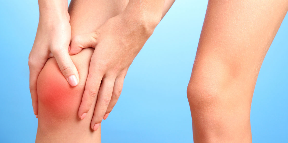
Pes anserine tendinopathy is a painful condition that affects the tendons on the inside of the knee. These tendons connect to the tibia bone, and their function is to allow flexion and stability of the knee. Pes anserine tendinopathy can cause significant pain and discomfort in this area.
Diagnosing pes anserine tendinopathy involves careful clinical evaluation and, in some cases, imaging tests.
The doctor will begin by obtaining a medical history that will include questions about the nature and duration of knee pain, activities that trigger or aggravate symptoms, and any previous injuries to the pes anserine area.
During the physical exam, the doctor will evaluate the knee for signs of tendinopathy. Functional tests can be performed to evaluate the strength and function of the muscles and tendons of the pes anserine area, as well as to determine the degree of impairment in mobility and the ability to perform daily activities.
In some cases, imaging tests, such as ultrasound or MRI, may be performed to evaluate the structure of the tendons and surrounding tissues.
Pes tendinopathy can present in a variety of ways, and patients may exhibit different phenotypes or patterns of presentation. Some of the most common presentations are described below:
- Acute Tendinopathy: Tendinopathy develops suddenly due to a traumatic injury or excessive strain. The pain can be intense and is accompanied by swelling and weakness in the knee.
- Chronic Tendinopathy: Chronic tendinopathy is usually the result of gradual wear and tear of the tendons due to repetitive activities, such as running or exercise. Symptoms may develop slowly over time and may include persistent pain, weakness, and limitation of function.
- Pes anserine bursitis: In some cases, tendinopathy may be associated with pes pes anserine bursitis, which involves inflammation of the synovial bursa in the area. This can cause swelling, pain, and limited mobility.
Treatment of pes tendinopathy depends on the specific phenotype, severity of the condition, and the individual needs of the patient. The most common treatment options are described below:
Treatment:
- Conservative management:
- Rest and Limitation of Activities: In acute cases, rest and limitation of activities that aggravate symptoms are essential.
- Physical Therapy: Strengthening and stretching exercises supervised by a physical therapist can help improve strength and flexibility in the affected area.
- Physical Therapy Modalities: Ultrasound therapy, shock wave therapy, and other physical therapy modalities can relieve pain and promote healing.
Pharmacological treatment:
Nonsteroidal anti-inflammatory drugs (NSAIDs) can relieve pain and reduce inflammation in mild to moderate cases.
In some moderate to severe cases, corticosteroid injections can be given directly to the affected area to successfully reduce inflammation and relieve pain.