

- Medical directory
- Specialities
- Specialised Units
 Teknon Cardiology Institute
 Obesity Unit
 Teknon Oncology Institute
 Bloodless Medicine and Surgery Unit
 Teknon Institute of Neurosciences
 Traffic Accident Care Unit
 Teknon Pulmonology Unit
 Maritime Medicine Unit
 Institute of Tissue Regenerative Therapy
 Teknon Pain Treatment Unit
 Teknon Tennis Clinic
 Systemic Inflammatory and Autoimmune Diseases Unit
 Assisted Reproduction Unit
 Sleep Unit
 Unidad de Síndromes de Sensibilización Central
- Specialised Units
- Diagnostics
- Diagnostic tests
 Diagnostic ImagingDiagnostic and interventional scans.
 Anatomical pathology laboratoryAllows to obtain a second opinion from renowned specialists.
 Clinical Analysis LaboratoryComprehensive service in the clinical area.
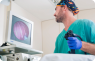 EndoscopyAn accurate diagnosis without conventional surgery.
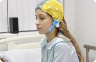 ElectrophysiologyFunctional exploration of the central nervous system.
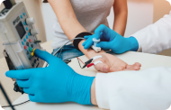 ElectromyographyClinical and neurophysiological evaluation of neuromuscular pathology.
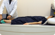 DensitometryDiagnostic technique for checking bone mineral density.
 UrodynamicsDiagnosis of urination disorders and incontinence.
- Medical check-ups

 GeneralThe most intelligent health check-up
 FullA comprehensive examination of your health
 Full PlusOur most exclusive check-up
 TravellersWhen travelling, your health is also part of your luggage
 SportA thorough review to boost your performance
 CardiologyGood news is knowing your heart is under control
 For companiesA tool that enhances employee satisfaction, productivity, and loyalty
- Diagnostic tests
- Our centre
- Teknon Healthcare Service Areas
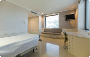 InpatientBright, functional and fully equipped rooms.
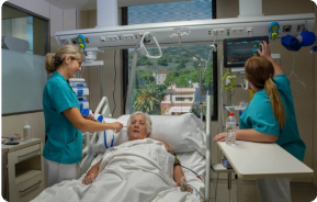 Semi-critical Care UnitEquipped with technology for diagnoses and treatments that require special care.
 Healthy Nutrition ProgrammeWe want to improve people’s health, which is why we promote healthy, conscious and sustainable nutrition at our hospitals.
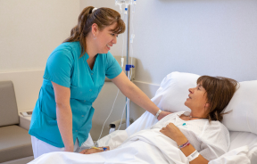 NursingOver 400 professionals.
 Emergency DepartmentUninterrupted operation, 24/7 at your service.
 Exclusivity / Teknon ClubCommitted to superior and individualised service, we furnish a full range of services in conjunction with our medical and healthcare support.
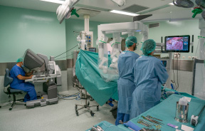 Surgical AreaA total of twenty (20) operating theatres, 12 of which are equipped for high-risk surgery.
 International ProgrammeA programme agent will provide you with comprehensive, personalised support
 Healthcare Ethics CommitteeGuidance for citizens and professionals in cases of moral conflicts.
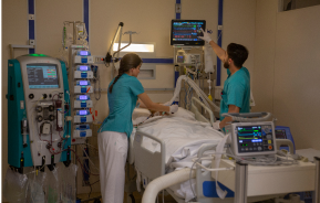 ICU-CCUMultipurpose unit incorporating treatment cubicles equipped with modern monitoring systems.
 Patient servicesAvailable to all our patients and their companions.
 ResearchResearch is one of the cornerstones of Centro Médico Teknon.
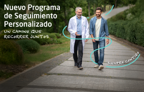 Personalised Follow-up ProgrammeWe accompany you throughout your medical journey. We organise your appointments and tests.
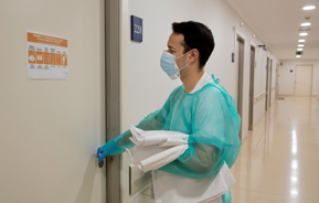 Patient Quality and SafetyOur management models are based on the most stringent national and international standards.
- Teknon Healthcare Service Areas
- News
- News
 NewsKeep abreast of the events at Centro Médico Teknon. Visit our News section.
 AgendaHere we post upcoming events and discussions on relevant health topics. Visit our Agenda section to see what’s up.
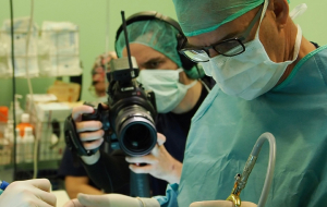 VideosThis section contains an extensive collection of videos related to our specialities.
 PodcastOur specialists discuss current medical topics, innovative treatments, health advice and patient experience.
 Health content
- News
- Blog

- Medical directory
- Specialities
- Specialised Units
- Teknon Cardiology Institute
- Obesity Unit
- Teknon Oncology Institute
- Bloodless Medicine and Surgery Unit
- Teknon Institute of Neurosciences
- Traffic Accident Care Unit
- Teknon Pulmonology Unit
- Maritime Medicine Unit
- Institute of Tissue Regenerative Therapy
- Teknon Pain Treatment Unit
- Teknon Tennis Clinic
- Systemic Inflammatory and Autoimmune Diseases Unit
- Assisted Reproduction Unit
- Sleep Unit
- Unidad de Síndromes de Sensibilización Central
- Specialised Units
- Diagnostics
- Diagnostic tests
- Diagnostic ImagingDiagnostic and interventional scans.
- Anatomical pathology laboratoryAllows to obtain a second opinion from renowned specialists.
- Clinical Analysis LaboratoryComprehensive service in the clinical area.
- EndoscopyAn accurate diagnosis without conventional surgery.
- ElectrophysiologyFunctional exploration of the central nervous system.
- ElectromyographyClinical and neurophysiological evaluation of neuromuscular pathology.
- DensitometryDiagnostic technique for checking bone mineral density.
- UrodynamicsDiagnosis of urination disorders and incontinence.
- Medical check-ups
- GeneralThe most intelligent health check-up
- FullA comprehensive examination of your health
- Full PlusOur most exclusive check-up
- TravellersWhen travelling, your health is also part of your luggage
- SportA thorough review to boost your performance
- CardiologyGood news is knowing your heart is under control
- For companiesA tool that enhances employee satisfaction, productivity, and loyalty
- Diagnostic tests
- Our centre
- Teknon Healthcare Service Areas
- InpatientBright, functional and fully equipped rooms.
- Semi-critical Care UnitEquipped with technology for diagnoses and treatments that require special care.
- Healthy Nutrition ProgrammeWe want to improve people’s health, which is why we promote healthy, conscious and sustainable nutrition at our hospitals.
- NursingOver 400 professionals.
- Emergency DepartmentUninterrupted operation, 24/7 at your service.
- Exclusivity / Teknon ClubCommitted to superior and individualised service, we furnish a full range of services in conjunction with our medical and healthcare support.
- Surgical AreaA total of twenty (20) operating theatres, 12 of which are equipped for high-risk surgery.
- International ProgrammeA programme agent will provide you with comprehensive, personalised support
- Healthcare Ethics CommitteeGuidance for citizens and professionals in cases of moral conflicts.
- ICU-CCUMultipurpose unit incorporating treatment cubicles equipped with modern monitoring systems.
- Patient servicesAvailable to all our patients and their companions.
- ResearchResearch is one of the cornerstones of Centro Médico Teknon.
- Personalised Follow-up ProgrammeWe accompany you throughout your medical journey. We organise your appointments and tests.
- Patient Quality and SafetyOur management models are based on the most stringent national and international standards.
- Teknon Healthcare Service Areas
- News
- News
- NewsKeep abreast of the events at Centro Médico Teknon. Visit our News section.
- AgendaHere we post upcoming events and discussions on relevant health topics. Visit our Agenda section to see what’s up.
- VideosThis section contains an extensive collection of videos related to our specialities.
- PodcastOur specialists discuss current medical topics, innovative treatments, health advice and patient experience.
- Health content
- News
- Blog


- Specialised Units
 Cardiology and Vascular Surgery
- Datos de interés
- State-of-the-art technology

State-of-the-art technology
Hybrid operating room: at the forefront of medical innovationThe team at the Teknon Cardiovascular Institute is expert in using the most advanced technology for the most accurate and reliable diagnosis of heart conditions and their treatment. We have a state-of-the-art hybrid operating room that allows us to perform minimally invasive procedures with the utmost safety and promoting rapid patient recovery.
Differences between a hybrid operating room and a conventional operating room
Benefits for patients
Less aggression for the patient, faster recovery after surgery, and less risk of infections and post-operative complications. These three factors are making minimally invasive surgery the standard for current surgical procedures.
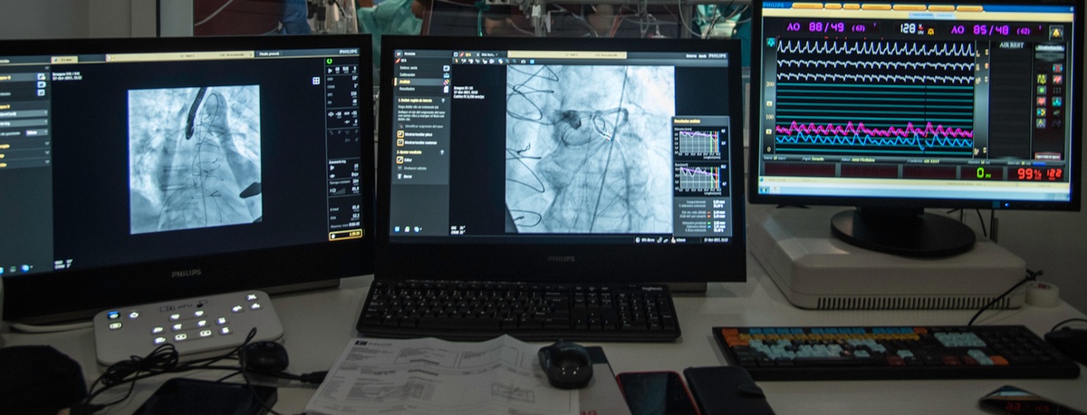
State-of-the-art haemodynamics rooms
Our goal is to offer the latest advances in cardiovascular disease treatment. We also have three haemodynamics and arrhythmia treatment rooms, each equipped with a state-of-the-art image-guided therapy platform. These platforms, with an intuitive interface similar to a tablet, allow complex procedures to be carried out in an app environment, working in parallel and scheduling large numbers of tasks in advance, which facilitates the work of professionals and increases patient safety.
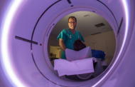
Advanced diagnostic imaging techniques
The most notable diagnostics techniques available are 3D echocardiography, cardiac magnetic resonance imaging, cardiac and coronary CAT scans, and PET CT scans.
- 3D echocardiography (3DE/3D Echo) 3D transthoracic echocardiography (3D-TTE) and transoesophageal echocardiography (3D-TEE) are accredited reference techniques for screening patients with heart valve disease. A simple ultrasound technique, 3D echocardiography is remarkable or rendering readily comprehensible images, making it the most lucid means to assess the heart, and an invaluable for cardiologists and other specialists treating heart disease patients. The most common indications for performing a 3D Echo examinations are:
- Aortic valve assessment: stenosis and regurgitation.
- Assessment of degenerative mitral valve with closure faults (insufficiency): mitral valve prolapse is one of the most common indications for 3D echocardiography, although it can also be useful for assessing mitral stenosis, especially in cases with poor transthoracic window.
- Prosthesis assessment: this is another of the main indications for the 3D-TTE and 3D-TEE. In particular, the assessment of periprosthetic dehiscence. The information is more accurate than 2D Echo.
- Assessment of the left atrium and left ear.
- Study of congenital heart disease.
- Guide to percutaneous interventional procedures.
- Assessment of intracardiac tumour masses.
- Cardiac Magnetic Resonance Imaging. Centro Médico Teknon is one of the few private centres that currently has a cardiac magnetic resonance imaging (MRI) machine, a state-of-the-art technique for evaluating patients with heart disease, and the most advanced technology for performing this procedure (high-field MRI machine, specific antenna for cardiac studies, advanced image reconstruction software, and CD recording and colour image printing).
 Coronary Multidetector CT (MDCT). The 64-slice coronary MDTC is a novel technique for viewing heart arteries with no need to insert catheters. It is recommended in patients with chest pain under investigation who are at intermediate risk of coronary heart disease, have atypical chest pain, or have had stress tests with inconclusive results. It consists of a helical scanner with high-speed rotation that allows images of the heart in motion to be acquired while the patient is moved inside the scanner.
Teknon also has a new-generation computed tomography scanner that can obtain an image of the heart in a quarter of a second, the time it takes for a single heartbeat, and can acquire images with very low doses of radiation, up to 82% less.
Computed Axial Tomography (CAT) angiography techniques are also essential in the study of the aortic valve, aorta, and peripheral arteries in patients who will undergo a transcatheter aortic valve implantation (TAVI) procedure without the need for surgery.- PET-CT. PET-CT systems provide a combination of metabolic information from PET (Positron Emission Tomography) images and morphological information from CT (Computed Tomography) images in a single scan. The images of the human body obtained using this technique are used to assess various diseases, including heart disease. It allows the blood flow to the heart to be determined and signs of coronary disease to be assessed. It is used to determine areas of reduced function that are alive and scar tissue caused by a previous heart attack. Together with myocardial perfusion imaging, PET scans can differentiate between non-functional heart muscle and viable heart muscle that could benefit from a procedure such as angioplasty or coronary bypass surgery to restore blood flow and improve heart function. It is also very useful in diagnosing infections of cardiac structures and valve prostheses.
Our team is at your complete disposal.
![]() Centro Médico Teknon
Centro Médico Teknon
Carrer de Vilana, 12, 08022 Barcelona
![]() Opening hours
Opening hours
Open 24 hours
![]() Phone Numbers
Phone Numbers
932 906 200
900 301 013