

- Medical directory
- Specialities
- Specialised Units
 Teknon Cardiology Institute
 Obesity Unit
 Teknon Oncology Institute
 Bloodless Medicine and Surgery Unit
 Teknon Institute of Neurosciences
 Traffic Accident Care Unit
 Teknon Pulmonology Unit
 Maritime Medicine Unit
 Institute of Tissue Regenerative Therapy
 Teknon Pain Treatment Unit
 Teknon Tennis Clinic
 Systemic Inflammatory and Autoimmune Diseases Unit
 Assisted Reproduction Unit
 Sleep Unit
 Unidad de Síndromes de Sensibilización Central
- Specialised Units
- Diagnostics
- Diagnostic tests
 Diagnostic ImagingDiagnostic and interventional scans.
 Anatomical pathology laboratoryAllows to obtain a second opinion from renowned specialists.
 Clinical Analysis LaboratoryComprehensive service in the clinical area.
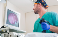 EndoscopyAn accurate diagnosis without conventional surgery.
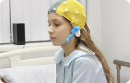 ElectrophysiologyFunctional exploration of the central nervous system.
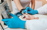 ElectromyographyClinical and neurophysiological evaluation of neuromuscular pathology.
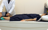 DensitometryDiagnostic technique for checking bone mineral density.
 UrodynamicsDiagnosis of urination disorders and incontinence.
- Medical check-ups

 GeneralThe most intelligent health check-up
 FullA comprehensive examination of your health
 Full PlusOur most exclusive check-up
 TravellersWhen travelling, your health is also part of your luggage
 SportA thorough review to boost your performance
 CardiologyGood news is knowing your heart is under control
 For companiesA tool that enhances employee satisfaction, productivity, and loyalty
- Diagnostic tests
- Our centre
- Teknon Healthcare Service Areas
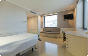 InpatientBright, functional and fully equipped rooms.
 Semi-critical Care UnitEquipped with technology for diagnoses and treatments that require special care.
 Healthy Nutrition ProgrammeWe want to improve people’s health, which is why we promote healthy, conscious and sustainable nutrition at our hospitals.
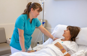 NursingOver 400 professionals.
 Emergency DepartmentUninterrupted operation, 24/7 at your service.
 Exclusivity / Teknon ClubCommitted to superior and individualised service, we furnish a full range of services in conjunction with our medical and healthcare support.
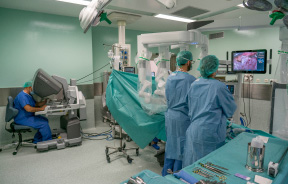 Surgical AreaA total of twenty (20) operating theatres, 12 of which are equipped for high-risk surgery.
 International ProgrammeA programme agent will provide you with comprehensive, personalised support
 Healthcare Ethics CommitteeGuidance for citizens and professionals in cases of moral conflicts.
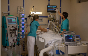 ICU-CCUMultipurpose unit incorporating treatment cubicles equipped with modern monitoring systems.
 Patient servicesAvailable to all our patients and their companions.
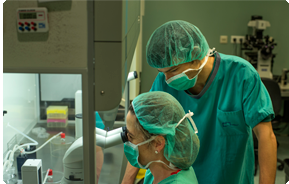 ResearchResearch is one of the cornerstones of Centro Médico Teknon.
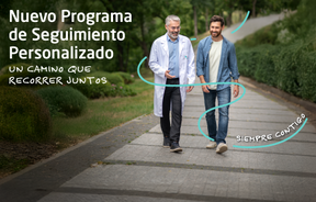 Personalised Follow-up ProgrammeWe accompany you throughout your medical journey. We organise your appointments and tests.
 Patient Quality and SafetyOur management models are based on the most stringent national and international standards.
- Teknon Healthcare Service Areas
- News
- News
 NewsKeep abreast of the events at Centro Médico Teknon. Visit our News section.
 AgendaHere we post upcoming events and discussions on relevant health topics. Visit our Agenda section to see what’s up.
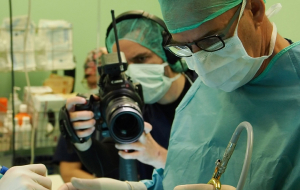 VideosThis section contains an extensive collection of videos related to our specialities.
 PodcastOur specialists discuss current medical topics, innovative treatments, health advice and patient experience.
 Health content
- News
- Blog

- Medical directory
- Specialities
- Specialised Units
- Teknon Cardiology Institute
- Obesity Unit
- Teknon Oncology Institute
- Bloodless Medicine and Surgery Unit
- Teknon Institute of Neurosciences
- Traffic Accident Care Unit
- Teknon Pulmonology Unit
- Maritime Medicine Unit
- Institute of Tissue Regenerative Therapy
- Teknon Pain Treatment Unit
- Teknon Tennis Clinic
- Systemic Inflammatory and Autoimmune Diseases Unit
- Assisted Reproduction Unit
- Sleep Unit
- Unidad de Síndromes de Sensibilización Central
- Specialised Units
- Diagnostics
- Diagnostic tests
- Diagnostic ImagingDiagnostic and interventional scans.
- Anatomical pathology laboratoryAllows to obtain a second opinion from renowned specialists.
- Clinical Analysis LaboratoryComprehensive service in the clinical area.
- EndoscopyAn accurate diagnosis without conventional surgery.
- ElectrophysiologyFunctional exploration of the central nervous system.
- ElectromyographyClinical and neurophysiological evaluation of neuromuscular pathology.
- DensitometryDiagnostic technique for checking bone mineral density.
- UrodynamicsDiagnosis of urination disorders and incontinence.
- Medical check-ups
- GeneralThe most intelligent health check-up
- FullA comprehensive examination of your health
- Full PlusOur most exclusive check-up
- TravellersWhen travelling, your health is also part of your luggage
- SportA thorough review to boost your performance
- CardiologyGood news is knowing your heart is under control
- For companiesA tool that enhances employee satisfaction, productivity, and loyalty
- Diagnostic tests
- Our centre
- Teknon Healthcare Service Areas
- InpatientBright, functional and fully equipped rooms.
- Semi-critical Care UnitEquipped with technology for diagnoses and treatments that require special care.
- Healthy Nutrition ProgrammeWe want to improve people’s health, which is why we promote healthy, conscious and sustainable nutrition at our hospitals.
- NursingOver 400 professionals.
- Emergency DepartmentUninterrupted operation, 24/7 at your service.
- Exclusivity / Teknon ClubCommitted to superior and individualised service, we furnish a full range of services in conjunction with our medical and healthcare support.
- Surgical AreaA total of twenty (20) operating theatres, 12 of which are equipped for high-risk surgery.
- International ProgrammeA programme agent will provide you with comprehensive, personalised support
- Healthcare Ethics CommitteeGuidance for citizens and professionals in cases of moral conflicts.
- ICU-CCUMultipurpose unit incorporating treatment cubicles equipped with modern monitoring systems.
- Patient servicesAvailable to all our patients and their companions.
- ResearchResearch is one of the cornerstones of Centro Médico Teknon.
- Personalised Follow-up ProgrammeWe accompany you throughout your medical journey. We organise your appointments and tests.
- Patient Quality and SafetyOur management models are based on the most stringent national and international standards.
- Teknon Healthcare Service Areas
- News
- News
- NewsKeep abreast of the events at Centro Médico Teknon. Visit our News section.
- AgendaHere we post upcoming events and discussions on relevant health topics. Visit our Agenda section to see what’s up.
- VideosThis section contains an extensive collection of videos related to our specialities.
- PodcastOur specialists discuss current medical topics, innovative treatments, health advice and patient experience.
- Health content
- News
- Blog


- Specialised Units
 Assisted Reproduction Unit
- Treatments and Specialities
- Female infertility study

Female infertility study
Commencement of the process
For women, the infertility study begins with:
- Hormonal and immunological analysis
It should be performed between the 2nd and 4th day of menstruation. There is no need to fast or skip meals.
 Hormonal: Follicle-Stimulating Hormone (FSH), Luteinizing Hormone (LH), Prolactin (PRL), and 17β-oestradiol, often alongside Anti-Müllerian Hormone (AMH).
- Immunological: HBsAg / HC Ac / RPR / Ac.VIH
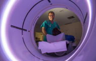
- Gynaecological ultrasound
The best time to perform an ultrasound scan is during the first few days of the cycle after menstruation. At this point, the follicular reserve of the ovaries can be assessed, anatomical abnormalities of the uterus (intrauterine deformities or septa) can be ruled out, endometrial alterations that hinder embryo implantation, such as the presence of endometrial polyps, can be identified, and ovarian cysts or tumours that may be causing infertility can be ruled out.
Based on these basic tests, the specialist will recommend additional diagnostic tests, either to identify other less likely causes of infertility or to investigate further any problems that may have been revealed in this basic study.
Additional tests for women
Hysterosalpingography (HSG)
Hysterosalpingography is performed when initial examinations are normal or when there is suspicion of possible tubal abnormalities, due to a history of endometriosis, previous surgery in women such as appendectomy or a history of peritonitis, etc. It also shows the correct morphology of the uterine cavity and its possible malformations: double, septal, arcuate uterus, etc. It is very useful for diagnosing dilations of the fallopian tubes such as hydrosalpinx, some of which can have negative effects on embryo implantation.
It is a simple technique that is performed at a radiology centre. Although it can sometimes be a little uncomfortable, it does not require anaesthesia. Performed under antibiotic prophylaxis, this technique has a very low complication rate (1%). It consists of introducing a radiopaque contrast fluid into the uterus through the vagina, which fills the uterine cavity and spreads through the fallopian tubes, outlining them and showing their permeability as it exits into the peritoneal cavity.
Hysteroscopy
Hysteroscopy involves inserting a thin camera into the uterus to view the inside of the uterine cavity. It allows for the assessment of internal alterations of the uterus, the presence of polyps and their removal, the presence of submucosal fibroids, etc.
It is a simple examination technique that does not always require anaesthesia.
Laparoscopy
Laparoscopy is a surgical examination technique performed in the operating room under general anaesthesia that allows access to the abdominal cavity to examine the correct morphology of the female reproductive system and correct any abnormalities. Sometimes the permeability of the fallopian tubes is also assessed, and pelvic adhesions that could alter the mobility of the tubes or trap the ovaries, making them difficult to access, are ruled out. It is a complementary invasive test and only performed if required by other infertility tests.
Karyotype
 Karyotyping involves taking a blood sample to determine each individual's chromosome pattern. Karyotype abnormalities may be linked to infertility problems or recurrent miscarriages. This entails an examination of 22 pairs of chromosomes and the 2 sex chromosomes: X and Y. Requested in cases of recurrent miscarriage, unexplained infertility, and sometimes before commencing an IVF-ICSI [In-Vitro Fertilization (IVF) - Intracytoplasmic Sperm Injection (ICSI)] cycle.
Infertility analysis (in cases of recurrent miscarriages)
To perform this analysis, a blood sample is taken and coagulation and immunological factors that may explain infertility are determined.