

- Medical directory
- Specialities
- Specialised Units
 Teknon Cardiology Institute
 Obesity Unit
 Teknon Oncology Institute
 Bloodless Medicine and Surgery Unit
 Teknon Institute of Neurosciences
 Traffic Accident Care Unit
 Teknon Pulmonology Unit
 Maritime Medicine Unit
 Institute of Tissue Regenerative Therapy
 Teknon Pain Treatment Unit
 Teknon Tennis Clinic
 Systemic Inflammatory and Autoimmune Diseases Unit
 Assisted Reproduction Unit
 Sleep Unit
 Unidad de Síndromes de Sensibilización Central
- Specialised Units
- Diagnostics
- Diagnostic tests
 Diagnostic ImagingDiagnostic and interventional scans.
 Anatomical pathology laboratoryAllows to obtain a second opinion from renowned specialists.
 Clinical Analysis LaboratoryComprehensive service in the clinical area.
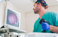 EndoscopyAn accurate diagnosis without conventional surgery.
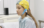 ElectrophysiologyFunctional exploration of the central nervous system.
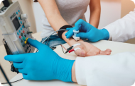 ElectromyographyClinical and neurophysiological evaluation of neuromuscular pathology.
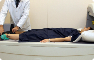 DensitometryDiagnostic technique for checking bone mineral density.
 UrodynamicsDiagnosis of urination disorders and incontinence.
- Medical check-ups

 GeneralThe most intelligent health check-up
 FullA comprehensive examination of your health
 Full PlusOur most exclusive check-up
 TravellersWhen travelling, your health is also part of your luggage
 SportA thorough review to boost your performance
 CardiologyGood news is knowing your heart is under control
 For companiesA tool that enhances employee satisfaction, productivity, and loyalty
- Diagnostic tests
- Our centre
- Teknon Healthcare Service Areas
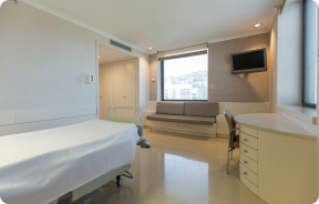 InpatientBright, functional and fully equipped rooms.
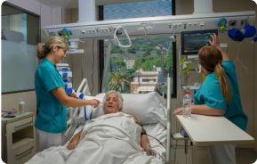 Semi-critical Care UnitEquipped with technology for diagnoses and treatments that require special care.
 Healthy Nutrition ProgrammeWe want to improve people’s health, which is why we promote healthy, conscious and sustainable nutrition at our hospitals.
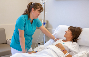 NursingOver 400 professionals.
 Emergency DepartmentUninterrupted operation, 24/7 at your service.
 Exclusivity / Teknon ClubCommitted to superior and individualised service, we furnish a full range of services in conjunction with our medical and healthcare support.
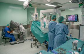 Surgical AreaA total of twenty (20) operating theatres, 12 of which are equipped for high-risk surgery.
 International ProgrammeA programme agent will provide you with comprehensive, personalised support
 Healthcare Ethics CommitteeGuidance for citizens and professionals in cases of moral conflicts.
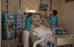 ICU-CCUMultipurpose unit incorporating treatment cubicles equipped with modern monitoring systems.
 Patient servicesAvailable to all our patients and their companions.
 ResearchResearch is one of the cornerstones of Centro Médico Teknon.
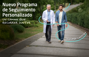 Personalised Follow-up ProgrammeWe accompany you throughout your medical journey. We organise your appointments and tests.
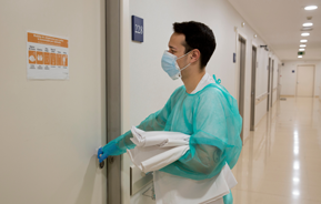 Patient Quality and SafetyOur management models are based on the most stringent national and international standards.
- Teknon Healthcare Service Areas
- News
- News
 NewsKeep abreast of the events at Centro Médico Teknon. Visit our News section.
 AgendaHere we post upcoming events and discussions on relevant health topics. Visit our Agenda section to see what’s up.
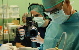 VideosThis section contains an extensive collection of videos related to our specialities.
 PodcastOur specialists discuss current medical topics, innovative treatments, health advice and patient experience.
 Health content
- News
- Blog

- Medical directory
- Specialities
- Specialised Units
- Teknon Cardiology Institute
- Obesity Unit
- Teknon Oncology Institute
- Bloodless Medicine and Surgery Unit
- Teknon Institute of Neurosciences
- Traffic Accident Care Unit
- Teknon Pulmonology Unit
- Maritime Medicine Unit
- Institute of Tissue Regenerative Therapy
- Teknon Pain Treatment Unit
- Teknon Tennis Clinic
- Systemic Inflammatory and Autoimmune Diseases Unit
- Assisted Reproduction Unit
- Sleep Unit
- Unidad de Síndromes de Sensibilización Central
- Specialised Units
- Diagnostics
- Diagnostic tests
- Diagnostic ImagingDiagnostic and interventional scans.
- Anatomical pathology laboratoryAllows to obtain a second opinion from renowned specialists.
- Clinical Analysis LaboratoryComprehensive service in the clinical area.
- EndoscopyAn accurate diagnosis without conventional surgery.
- ElectrophysiologyFunctional exploration of the central nervous system.
- ElectromyographyClinical and neurophysiological evaluation of neuromuscular pathology.
- DensitometryDiagnostic technique for checking bone mineral density.
- UrodynamicsDiagnosis of urination disorders and incontinence.
- Medical check-ups
- GeneralThe most intelligent health check-up
- FullA comprehensive examination of your health
- Full PlusOur most exclusive check-up
- TravellersWhen travelling, your health is also part of your luggage
- SportA thorough review to boost your performance
- CardiologyGood news is knowing your heart is under control
- For companiesA tool that enhances employee satisfaction, productivity, and loyalty
- Diagnostic tests
- Our centre
- Teknon Healthcare Service Areas
- InpatientBright, functional and fully equipped rooms.
- Semi-critical Care UnitEquipped with technology for diagnoses and treatments that require special care.
- Healthy Nutrition ProgrammeWe want to improve people’s health, which is why we promote healthy, conscious and sustainable nutrition at our hospitals.
- NursingOver 400 professionals.
- Emergency DepartmentUninterrupted operation, 24/7 at your service.
- Exclusivity / Teknon ClubCommitted to superior and individualised service, we furnish a full range of services in conjunction with our medical and healthcare support.
- Surgical AreaA total of twenty (20) operating theatres, 12 of which are equipped for high-risk surgery.
- International ProgrammeA programme agent will provide you with comprehensive, personalised support
- Healthcare Ethics CommitteeGuidance for citizens and professionals in cases of moral conflicts.
- ICU-CCUMultipurpose unit incorporating treatment cubicles equipped with modern monitoring systems.
- Patient servicesAvailable to all our patients and their companions.
- ResearchResearch is one of the cornerstones of Centro Médico Teknon.
- Personalised Follow-up ProgrammeWe accompany you throughout your medical journey. We organise your appointments and tests.
- Patient Quality and SafetyOur management models are based on the most stringent national and international standards.
- Teknon Healthcare Service Areas
- News
- News
- NewsKeep abreast of the events at Centro Médico Teknon. Visit our News section.
- AgendaHere we post upcoming events and discussions on relevant health topics. Visit our Agenda section to see what’s up.
- VideosThis section contains an extensive collection of videos related to our specialities.
- PodcastOur specialists discuss current medical topics, innovative treatments, health advice and patient experience.
- Health content
- News
- Blog


- Specialised Units
 Diagnostic tests
- Treatments and Specialities
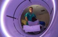
- Diagnostic Imaging
- Digitalised conventional radiology
Digitalised conventional radiology
Digitalised Conventional Radiology facilitates the observation of the internal structures of the body to study possible lesions. New techniques now render the use of remarkably reduced levels of ionising radiation viable, which subsequently simplifies the process and procures sharper and higher quality images to incorporate the data into the centre’s computer system.
- Neuroradiology
- Skull X-ray

This technique uses X-ray rendered imaging for examining the skull. Indicated for: trauma, premature cranial suture closure.
- Paranasal Sinuses X-ray

This technique uses X-ray rendered imaging for examining the paranasal sinuses. Indicated for: difficulty breathing through the nose, chronic cough, headache, mucus.
- TMJ X-ray (Temporomandibular Joint)

This technique uses X-ray rendered imaging for examining the temporomandibular joint. Indicated for: trauma, chewing pain, chewing noise, limited mouth opening.
- Cavum X-ray

This technique uses X-ray rendered imaging for examining the cavum. Indicated for: respiratory distress, recurrent angina in infants and children.
- Cervical spine X-ray

This technique uses X-ray rendered imaging for examining the cervical spine. Indicated for: trauma, cervical contracture, joint pain.
- Nasal bone anatomy X-ray

This technique uses X-ray rendered imaging for examining the nasal bone anatomy. Indicated for: trauma, plastic surgery planning.
- Internal auditory canal X-ray

This technique uses X-ray rendered imaging for examining the internal auditory canal. Indicated for: hearing loss.
- Orbit X-ray

This technique uses X-ray rendered imaging for examining the orbits. Indicated for: ocular foreign body, trauma, infections.
- Mandibular X-ray

This technique uses X-ray rendered imaging for examining the jaw. Indicated for: trauma, congenital anomalies.
- Laryngeal X-ray

This technique uses X-ray rendered imaging for examining the larynx. Indicated for: respiratory distress.
- Neck X-ray, soft tissue

This technique uses X-ray rendered imaging for examining the neck. Indicated for: study of lumps or nodules.
- Cranial base X-ray

This technique uses X-ray rendered imaging for examining the cranial base. Indicated for: study of congenital malformations.
- Thorax
- Chest X-ray

This technique uses X-ray rendered imaging for examining the thoracic cavity (heart, lungs, costal arches, clavicles, etc.).
- Sternum X-ray
- Clavicle X-ray
- Costal chest X-ray
- Thoracic spine X-ray

This technique uses X-ray rendered imaging for examining the thoracic spine. Indicated for: trauma, pain, scoliosis.
- Oesophagogram

Diagnostic test to obtain moving radiological images of the oesophagus, using X-rays (fluoroscopy) and an opaque barium contrast agent administered orally.
- Abdomen and pelvis
- Abdomen X-ray

This technique uses X-ray rendered imaging for examining the abdomen (stomach, small intestine, large intestine, liver, kidneys, bladder, bony pelvis, etc.).
- Lumbar spine X-ray
- EGDT (Oesophageal-gastro-duodenal transit)

Diagnostic test to obtain moving radiological images of the oesophagus, stomach and duodenum using X-rays (fluoroscopy) and an opaque barium contrast agent administered orally.
- Intestinal transit

Diagnostic test to obtain moving radiological images of the oesophagus, stomach, duodenum and small intestine using X-rays (fluoroscopy) and an opaque barium contrast agent administered orally.
- Double-contrast barium enema
X-ray based diagnostic testing to obtain radiological images of the large intestine (colon and rectum). An opaque contrast agent is administered through the rectum in the form of an enema, and air is also introduced to expand the colon.
- IVU (Retrograde pyelography)

Intravenous urography (IVU) involves serial radiological imaging of the kidney, urinary tract and bladder. This study always requires the use of an iodinated contrast agent.
- Voiding Cystourethrography (VCUG)

Serial voiding cystourethrography involves radiological imaging to assess the anatomy and function of the urethra and bladder with the administration of iodinated contrast through a bladder catheter.
- Urethrocystography (Urethro-CG)

Retrograde-minctional urethrocystrography involves radiological imaging to assess the anatomy and function of the bladder and urethra. Contrast material is applied through a small catheter placed in the urethra and images are obtained during the filling and emptying of the bladder.
- Ascending catheter pyelography
Diagnostic test to visualise the ureter and renal pelvis using X-rays in patients with a catheter by injecting an iodinated contrast agent through the catheter.
- Nephrostomy pyelography
Diagnostic test to visualise the urinary tract using X-rays in patients with a nephrostomy catheter by injecting an iodinated contrast agent through the catheter.
- Hypsterosalpingography (HSG)

Diagnostic test to visualise and study the uterus and fallopian tubes using X-rays and a contrast medium. The patient should be given prophylactic antibiotic treatment. It is recommended that a mild sedative be taken one hour before the test.
- Trans-Kher Cholangiography
Diagnostic test to visualise the biliary tract using X-rays in patients with a Kehr tube by injecting an iodinated contrast agent through the tube.
- Osteoarticular
- Limb X-ray

This technique uses X-ray imaging to examine the limbs, especially the bones. Indicated for: trauma, joint infections.
- X-ray arthrography

This X-ray based procedure involves injecting a contrast agent into the joint to detect lesions in certain structures (cartilage, bone, tendons, etc.) by observing the distribution of the contrast agent.
- Pelvis X-ray

This procedure uses X-ray imaging to examine the pelvis, especially the pelvic bones.
- Bone age X-ray

X-ray examination of the hand to assess the patient’s age based on the development of the bones and to determine if there is a delay or growth greater than that corresponding to the patient’s age.
- Paediatric hip X-ray
- Serial bone X-ray

X-ray screening of different body structures (skull, spine, ribs, pelvis, humerus and femurs) to rule out bone injuries. Indicated for: study of multiple myeloma, monoclonal gammopathies, metastases.
- Sacroiliac X-ray

This procedure uses X-rays to examine the sacroiliac joints and rule out inflammatory, traumatic or degenerative diseases.
- Spinal column
- Cervical spine X-ray

This technique uses X-ray rendered imaging for examining the cervical spine. Indicated for: trauma, cervical pain.
- Thoracic spine X-ray

This technique uses X-ray rendered imaging to examine the thoracic spine. Indicated for: trauma, back pain.
- Lumbar spine X-ray

This technique uses X-ray rendered imaging to examine the lumbar spine. Indicated for: sciatica, trauma, lower back pain.
- Sacrum-coccyx X-ray

This technique uses X-ray rendered imaging to examine the sacrum and coccyx. Indicated for: trauma, sacral or coccygeal pain.
- Spinal teleradiology

This technique uses X-ray rendered imaging to examine the entire spinal column, especially assessing the presence of scoliosis and pelvic dysmetria.
- Paediatrics
- Cavum X-ray

This technique uses X-ray rendered imaging for examining the cavum. Indicated for: respiratory distress, regurgitation, recurrent otitis.
- Bone age X-ray

X-ray examination of the hand to assess the patient’s age based on the development of the bones and to determine if there is a delay or growth greater than that corresponding to the patient’s age.
- Paranasal Sinuses X-ray

This technique uses X-ray rendered imaging for examining the paranasal sinuses. Indicated for: headache, chronic cough.
- Chest X-ray

A chest X-ray uses a small dose of radiation to obtain a two-dimensional image of the thoracic cavity and its contents (lungs, heart, mediastinum, etc.). It is one of the most commonly performed medical tests.
- Abdomen X-ray

An abdominal X-ray uses a small dose of radiation to obtain a two-dimensional image of the abdomen with its anatomical structures (stomach, small intestine, large intestine, liver, pancreas, kidneys, bladder, bony pelvis, etc.).
- Spinal teleradiology

This technique uses X-ray rendered imaging to examine the entire spinal column, especially assessing the presence of scoliosis and pelvic dysmetria.
- Telemetry (Lower Limbs)

This technique uses X-ray rendered imaging to examine both lower limbs in their entirety, especially assessing the presence of dysmetria between the two.
- Limb X-ray
- Hip X-ray
- Cystography

Serial voiding cystourethrography involves radiological imaging to assess the anatomy and function of the urethra and bladder with the administration of iodinated contrast through a bladder catheter.


