

- Medical directory
- Specialities
- Specialised Units
 Teknon Cardiology Institute
 Obesity Unit
 Teknon Oncology Institute
 Bloodless Medicine and Surgery Unit
 Teknon Institute of Neurosciences
 Traffic Accident Care Unit
 Teknon Pulmonology Unit
 Maritime Medicine Unit
 Institute of Tissue Regenerative Therapy
 Teknon Pain Treatment Unit
 Teknon Tennis Clinic
 Systemic Inflammatory and Autoimmune Diseases Unit
 Assisted Reproduction Unit
 Sleep Unit
 Unidad de Síndromes de Sensibilización Central
- Specialised Units
- Diagnostics
- Diagnostic tests
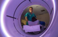 Diagnostic ImagingDiagnostic and interventional scans.
 Anatomical pathology laboratoryAllows to obtain a second opinion from renowned specialists.
 Clinical Analysis LaboratoryComprehensive service in the clinical area.
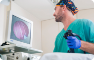 EndoscopyAn accurate diagnosis without conventional surgery.
 ElectrophysiologyFunctional exploration of the central nervous system.
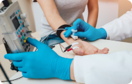 ElectromyographyClinical and neurophysiological evaluation of neuromuscular pathology.
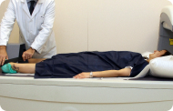 DensitometryDiagnostic technique for checking bone mineral density.
 UrodynamicsDiagnosis of urination disorders and incontinence.
- Medical check-ups

 GeneralThe most intelligent health check-up
 FullA comprehensive examination of your health
 Full PlusOur most exclusive check-up
 TravellersWhen travelling, your health is also part of your luggage
 SportA thorough review to boost your performance
 CardiologyGood news is knowing your heart is under control
 For companiesA tool that enhances employee satisfaction, productivity, and loyalty
- Diagnostic tests
- Our centre
- Teknon Healthcare Service Areas
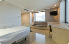 InpatientBright, functional and fully equipped rooms.
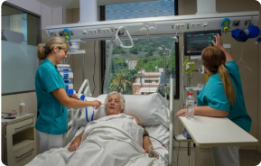 Semi-critical Care UnitEquipped with technology for diagnoses and treatments that require special care.
 Healthy Nutrition ProgrammeWe want to improve people’s health, which is why we promote healthy, conscious and sustainable nutrition at our hospitals.
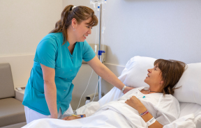 NursingOver 400 professionals.
 Emergency DepartmentUninterrupted operation, 24/7 at your service.
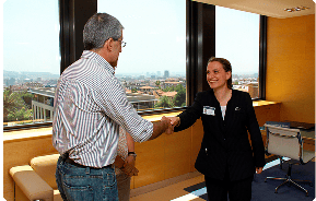 Exclusivity / Teknon ClubCommitted to superior and individualised service, we furnish a full range of services in conjunction with our medical and healthcare support.
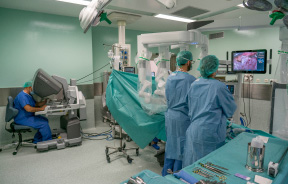 Surgical AreaA total of twenty (20) operating theatres, 12 of which are equipped for high-risk surgery.
 International ProgrammeA programme agent will provide you with comprehensive, personalised support
 Healthcare Ethics CommitteeGuidance for citizens and professionals in cases of moral conflicts.
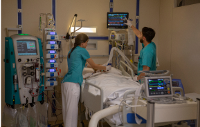 ICU-CCUMultipurpose unit incorporating treatment cubicles equipped with modern monitoring systems.
 Patient servicesAvailable to all our patients and their companions.
 ResearchResearch is one of the cornerstones of Centro Médico Teknon.
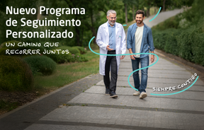 Personalised Follow-up ProgrammeWe accompany you throughout your medical journey. We organise your appointments and tests.
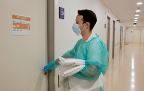 Patient Quality and SafetyOur management models are based on the most stringent national and international standards.
- Teknon Healthcare Service Areas
- News
- News
 NewsKeep abreast of the events at Centro Médico Teknon. Visit our News section.
 AgendaHere we post upcoming events and discussions on relevant health topics. Visit our Agenda section to see what’s up.
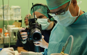 VideosThis section contains an extensive collection of videos related to our specialities.
 PodcastOur specialists discuss current medical topics, innovative treatments, health advice and patient experience.
 Health content
- News
- Blog

- Medical directory
- Specialities
- Specialised Units
- Teknon Cardiology Institute
- Obesity Unit
- Teknon Oncology Institute
- Bloodless Medicine and Surgery Unit
- Teknon Institute of Neurosciences
- Traffic Accident Care Unit
- Teknon Pulmonology Unit
- Maritime Medicine Unit
- Institute of Tissue Regenerative Therapy
- Teknon Pain Treatment Unit
- Teknon Tennis Clinic
- Systemic Inflammatory and Autoimmune Diseases Unit
- Assisted Reproduction Unit
- Sleep Unit
- Unidad de Síndromes de Sensibilización Central
- Specialised Units
- Diagnostics
- Diagnostic tests
- Diagnostic ImagingDiagnostic and interventional scans.
- Anatomical pathology laboratoryAllows to obtain a second opinion from renowned specialists.
- Clinical Analysis LaboratoryComprehensive service in the clinical area.
- EndoscopyAn accurate diagnosis without conventional surgery.
- ElectrophysiologyFunctional exploration of the central nervous system.
- ElectromyographyClinical and neurophysiological evaluation of neuromuscular pathology.
- DensitometryDiagnostic technique for checking bone mineral density.
- UrodynamicsDiagnosis of urination disorders and incontinence.
- Medical check-ups
- GeneralThe most intelligent health check-up
- FullA comprehensive examination of your health
- Full PlusOur most exclusive check-up
- TravellersWhen travelling, your health is also part of your luggage
- SportA thorough review to boost your performance
- CardiologyGood news is knowing your heart is under control
- For companiesA tool that enhances employee satisfaction, productivity, and loyalty
- Diagnostic tests
- Our centre
- Teknon Healthcare Service Areas
- InpatientBright, functional and fully equipped rooms.
- Semi-critical Care UnitEquipped with technology for diagnoses and treatments that require special care.
- Healthy Nutrition ProgrammeWe want to improve people’s health, which is why we promote healthy, conscious and sustainable nutrition at our hospitals.
- NursingOver 400 professionals.
- Emergency DepartmentUninterrupted operation, 24/7 at your service.
- Exclusivity / Teknon ClubCommitted to superior and individualised service, we furnish a full range of services in conjunction with our medical and healthcare support.
- Surgical AreaA total of twenty (20) operating theatres, 12 of which are equipped for high-risk surgery.
- International ProgrammeA programme agent will provide you with comprehensive, personalised support
- Healthcare Ethics CommitteeGuidance for citizens and professionals in cases of moral conflicts.
- ICU-CCUMultipurpose unit incorporating treatment cubicles equipped with modern monitoring systems.
- Patient servicesAvailable to all our patients and their companions.
- ResearchResearch is one of the cornerstones of Centro Médico Teknon.
- Personalised Follow-up ProgrammeWe accompany you throughout your medical journey. We organise your appointments and tests.
- Patient Quality and SafetyOur management models are based on the most stringent national and international standards.
- Teknon Healthcare Service Areas
- News
- News
- NewsKeep abreast of the events at Centro Médico Teknon. Visit our News section.
- AgendaHere we post upcoming events and discussions on relevant health topics. Visit our Agenda section to see what’s up.
- VideosThis section contains an extensive collection of videos related to our specialities.
- PodcastOur specialists discuss current medical topics, innovative treatments, health advice and patient experience.
- Health content
- News
- Blog


- Specialised Units
 Diagnostic tests
- Treatments and Specialities
- Diagnostic Imaging
- Mammography
Mammography
Mammography tests use low-dose x-ray imaging to detect breast tissue pathologies (especially breast cancer) in gynaecological screening and targeted diagnostics. Mammography can be interventional with cyst puncture, stereotactic biopsy, pre-surgical marking, and galactography.
Digital mammography

Digital mammography is an innovative breast examination technique that uses a low radiation dose, thus reducing exposure compared to conventional mammography. The image is digitised using special cassettes on a system known as CR (Computed Radiography). These digitalisation techniques provide excellent images, especially in breasts that are difficult to examine, such as glandular breasts, which appear dense on mammograms, enabling better detection of tumours in this group of patients.
Mammography-guided stereotactic biopsy

The presence of clustered microcalcifications may be an early form of breast cancer. Stereotactic biopsy is used to diagnose these small lesions. In this procedure, the lesion is located on the mammogram and a computer connected to the mammography machine uses a guidance system to determine the exact point and depth of the puncture. The radiologist performs the biopsy after administering local anaesthesia to the area, removing several millimetre-sized fragments of the lesion (5 to 10) so that the pathologist can then diagnose the condition. The average duration of the procedure is 20 minutes and causes slight discomfort.
Pre-surgical harpoon marking

Small, non-palpable breast lesions requiring limited surgery, which need to be marked prior to the procedure by placing a harpoon in the area to be examined. The harpoon is a thin wire with an angled tip (harpoon) that remains fixed in the designated area of the breast. Under mammographic or ultrasound guidance, the radiologist inserts the harpoon into the lesion, where it remains fixed. This will enable the surgeon to locate the injury safely. The average duration of the procedure is 20 minutes and causes slight discomfort. It is usually performed a few hours before going into the operating theatre, although it can be done the day before.
Breast ultrasound

This imaging method uses sound waves create an image of blood vessels, tissues and organs, including the breasts. Ultrasound imaging allows for the evaluation of abnormalities detected by mammography or clinical examination, distinguishing between solid and fluid lesions (cysts).
Ultrasound-guided FNA (fine-needle aspiration)

In fine needle aspiration (FNA), a thin needle is inserted under ultrasound guidance into a suspicious area of the breast; a syringe is then used to extract a small amount of tissue, which is sent for cytological examination. It is also used to puncture and drain breast cysts and analyse their contents. The procedure lasts only a few minutes and causes slight discomfort.
Ultrasound-guided CNB (core-needle biopsy)
A core-needle biopsy or core biopsy involves obtaining a cylinder of tissue for histological study. Under ultrasound guidance and after administering local anaesthesia, the needle is placed in the area to be studied and, using an automatic cutting system, several millimetre-long cylinders of tissue are obtained. It is a relatively short and relatively painless procedure. It allows a significant sample to be obtained for histological study.
Gold seeding for radiotherapy
It consists of marking diagnosed cancer lesions to indicate their exact location in order to apply radiotherapy selectively so that it affects the tumour with minimal impact on neighbouring tissues. This procedure is performed under ultrasound guidance, locating the tumour and inserting the gold seeds (3 to 4 seeds) as markers at four points around its periphery.
Pre-chemotherapy titanium clipping

Chemotherapy may be administered prior to surgery in the treatment of breast cancer. The tumours decrease in size, sometimes very markedly, making them difficult to locate during surgery. The technique involves placing a titanium clip in the centre of the lesions diagnosed as cancerous. It is performed under ultrasound guidance in most cases, or by mammography.
Galactography (ductography)

This radiological technique examines the mammary ducts to diagnose the presence of lesions inside them, usually small tumours or stenosis. It is indicated in patients who present nipple discharge when it affects only a single nipple ductal orifice. The technique consists of introducing contrast, usually about 2 cc, into the opening of the inadequate secretion, prior to dilating it. A series of mammograms are then performed to study the duct. It is a thorough but painless test.