

- Medical directory
- Specialities
- Specialised Units
 Teknon Cardiology Institute
 Obesity Unit
 Teknon Oncology Institute
 Bloodless Medicine and Surgery Unit
 Teknon Institute of Neurosciences
 Traffic Accident Care Unit
 Teknon Pulmonology Unit
 Maritime Medicine Unit
 Institute of Tissue Regenerative Therapy
 Teknon Pain Treatment Unit
 Teknon Tennis Clinic
 Systemic Inflammatory and Autoimmune Diseases Unit
 Assisted Reproduction Unit
 Sleep Unit
 Unidad de Síndromes de Sensibilización Central
- Specialised Units
- Diagnostics
- Diagnostic tests
 Diagnostic ImagingDiagnostic and interventional scans.
 Anatomical pathology laboratoryAllows to obtain a second opinion from renowned specialists.
 Clinical Analysis LaboratoryComprehensive service in the clinical area.
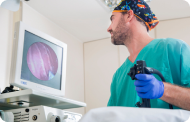 EndoscopyAn accurate diagnosis without conventional surgery.
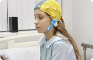 ElectrophysiologyFunctional exploration of the central nervous system.
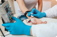 ElectromyographyClinical and neurophysiological evaluation of neuromuscular pathology.
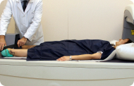 DensitometryDiagnostic technique for checking bone mineral density.
 UrodynamicsDiagnosis of urination disorders and incontinence.
- Medical check-ups

 GeneralThe most intelligent health check-up
 FullA comprehensive examination of your health
 Full PlusOur most exclusive check-up
 TravellersWhen travelling, your health is also part of your luggage
 SportA thorough review to boost your performance
 CardiologyGood news is knowing your heart is under control
 For companiesA tool that enhances employee satisfaction, productivity, and loyalty
- Diagnostic tests
- Our centre
- Teknon Healthcare Service Areas
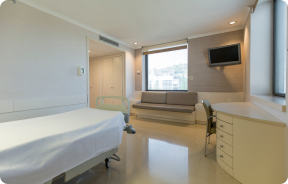 InpatientBright, functional and fully equipped rooms.
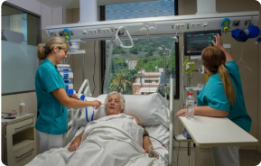 Semi-critical Care UnitEquipped with technology for diagnoses and treatments that require special care.
 Healthy Nutrition ProgrammeWe want to improve people’s health, which is why we promote healthy, conscious and sustainable nutrition at our hospitals.
 NursingOver 400 professionals.
 Emergency DepartmentUninterrupted operation, 24/7 at your service.
 Exclusivity / Teknon ClubCommitted to superior and individualised service, we furnish a full range of services in conjunction with our medical and healthcare support.
 Surgical AreaA total of twenty (20) operating theatres, 12 of which are equipped for high-risk surgery.
 International ProgrammeA programme agent will provide you with comprehensive, personalised support
 Healthcare Ethics CommitteeGuidance for citizens and professionals in cases of moral conflicts.
 ICU-CCUMultipurpose unit incorporating treatment cubicles equipped with modern monitoring systems.
 Patient servicesAvailable to all our patients and their companions.
 ResearchResearch is one of the cornerstones of Centro Médico Teknon.
 Personalised Follow-up ProgrammeWe accompany you throughout your medical journey. We organise your appointments and tests.
 Patient Quality and SafetyOur management models are based on the most stringent national and international standards.
- Teknon Healthcare Service Areas
- News
- News
 NewsKeep abreast of the events at Centro Médico Teknon. Visit our News section.
 AgendaHere we post upcoming events and discussions on relevant health topics. Visit our Agenda section to see what’s up.
 VideosThis section contains an extensive collection of videos related to our specialities.
 PodcastOur specialists discuss current medical topics, innovative treatments, health advice and patient experience.
 Health content
- News
- Blog

- Medical directory
- Specialities
- Specialised Units
- Teknon Cardiology Institute
- Obesity Unit
- Teknon Oncology Institute
- Bloodless Medicine and Surgery Unit
- Teknon Institute of Neurosciences
- Traffic Accident Care Unit
- Teknon Pulmonology Unit
- Maritime Medicine Unit
- Institute of Tissue Regenerative Therapy
- Teknon Pain Treatment Unit
- Teknon Tennis Clinic
- Systemic Inflammatory and Autoimmune Diseases Unit
- Assisted Reproduction Unit
- Sleep Unit
- Unidad de Síndromes de Sensibilización Central
- Specialised Units
- Diagnostics
- Diagnostic tests
- Diagnostic ImagingDiagnostic and interventional scans.
- Anatomical pathology laboratoryAllows to obtain a second opinion from renowned specialists.
- Clinical Analysis LaboratoryComprehensive service in the clinical area.
- EndoscopyAn accurate diagnosis without conventional surgery.
- ElectrophysiologyFunctional exploration of the central nervous system.
- ElectromyographyClinical and neurophysiological evaluation of neuromuscular pathology.
- DensitometryDiagnostic technique for checking bone mineral density.
- UrodynamicsDiagnosis of urination disorders and incontinence.
- Medical check-ups
- GeneralThe most intelligent health check-up
- FullA comprehensive examination of your health
- Full PlusOur most exclusive check-up
- TravellersWhen travelling, your health is also part of your luggage
- SportA thorough review to boost your performance
- CardiologyGood news is knowing your heart is under control
- For companiesA tool that enhances employee satisfaction, productivity, and loyalty
- Diagnostic tests
- Our centre
- Teknon Healthcare Service Areas
- InpatientBright, functional and fully equipped rooms.
- Semi-critical Care UnitEquipped with technology for diagnoses and treatments that require special care.
- Healthy Nutrition ProgrammeWe want to improve people’s health, which is why we promote healthy, conscious and sustainable nutrition at our hospitals.
- NursingOver 400 professionals.
- Emergency DepartmentUninterrupted operation, 24/7 at your service.
- Exclusivity / Teknon ClubCommitted to superior and individualised service, we furnish a full range of services in conjunction with our medical and healthcare support.
- Surgical AreaA total of twenty (20) operating theatres, 12 of which are equipped for high-risk surgery.
- International ProgrammeA programme agent will provide you with comprehensive, personalised support
- Healthcare Ethics CommitteeGuidance for citizens and professionals in cases of moral conflicts.
- ICU-CCUMultipurpose unit incorporating treatment cubicles equipped with modern monitoring systems.
- Patient servicesAvailable to all our patients and their companions.
- ResearchResearch is one of the cornerstones of Centro Médico Teknon.
- Personalised Follow-up ProgrammeWe accompany you throughout your medical journey. We organise your appointments and tests.
- Patient Quality and SafetyOur management models are based on the most stringent national and international standards.
- Teknon Healthcare Service Areas
- News
- News
- NewsKeep abreast of the events at Centro Médico Teknon. Visit our News section.
- AgendaHere we post upcoming events and discussions on relevant health topics. Visit our Agenda section to see what’s up.
- VideosThis section contains an extensive collection of videos related to our specialities.
- PodcastOur specialists discuss current medical topics, innovative treatments, health advice and patient experience.
- Health content
- News
- Blog


- Specialised Units
 Diagnostic tests
- Treatments and Specialities
- Diagnostic Imaging
- Magnetic Resonance Imaging
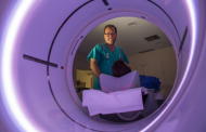
Magnetic Resonance Imaging
Magnetic resonance imaging (MRI) is a non-invasive diagnostic test (which does not use X-rays) that produces high-definition anatomical images based on the resonance produced in tissues subjected to a magnetic field. This imaging technique provides the greatest differentiation between different tissues in the body, allowing very accurate information to be obtained about the structure and composition of the organ being analysed.
- Centro Médico Teknon has the first high-field open MRI scanner in Catalonia. The device can also be used to examine patients not only in a lying position, but also with their joints flexed and in motion.
- Neuroradiology
- Brain MRI (cranial)

This non-invasive diagnostic procedure uses an electromagnetic field and radio waves (from a transmitter and receiver) to acquire high-definition anatomical images of the brain. It is a radiation-free procedure. Indicated for: vascular problems, memory loss, epilepsy, headache, malformations, suspected tumour, meningitis.
- IAC MRI

This non-invasive diagnostic procedure uses an electromagnetic field and radio waves (from a transmitter and receiver) to acquire high-definition anatomical images of the internal auditory canal (IAC). It is a radiation-free procedure. Indicated for: hearing problems, dizziness, vertigo.
- Neck MRI
This non-invasive diagnostic procedure uses an electromagnetic field and radio waves (from a transmitter and receiver) to acquire high-definition anatomical images of the neck. It is a radiation-free procedure. Indicated for: suspected tumours, infections, lymph nodes.
- Intracranial MRI angiography

This non-invasive diagnostic procedure uses an electromagnetic field and radio waves (from a transmitter and receiver) to acquire high-definition anatomical images of the cerebral arteries. It is a radiation-free procedure. In most cases, paramagnetic contrast (gadolinium) is required. It enables non-invasive angiographic studies using a gadolinium injection, with subsequent 2D and 3D reconstruction using specialised workstations. Indicated for: vascular malformations, cerebral artery aneurysms, arteriosclerosis.
- Supra-aortic trunk MRI angiography
This non-invasive diagnostic procedure uses an electromagnetic field and radio waves (from a transmitter and receiver) to acquire high-definition anatomical images of the carotid and vertebral arteries in the neck. It is a radiation-free procedure. In most cases, paramagnetic contrast (gadolinium) is required. It enables non-invasive angiographic studies using a gadolinium injection, with subsequent 2D and 3D reconstruction using specialised workstations. Indicated for: cerebral circulatory problems, syncope.
- Brain MRI spectroscopy
This non-invasive diagnostic procedure uses an electromagnetic field and radio waves (from a transmitter and receiver) to acquire high-definition anatomical images of the brain. It is a radiation-free procedure. This is followed by a qualitative and quantitative measurement of various metabolites (substances) to help characterise lesions. Indicated for: tumours, metabolic disorders, infections, epilepsy.
- Cerebrospinal fluid (CSF) flow MRI
This non-invasive diagnostic procedure uses an electromagnetic field and radio waves (from a transmitter and receiver) to acquire high-definition anatomical images of the cerebral ventricular system. It is a radiation-free procedure. Indicated for: study of hydrocephalus and stenosis of the cerebral aqueduct.
- Cervical spine MRI

This non-invasive diagnostic procedure uses an electromagnetic field and radio waves (from a transmitter and receiver) to acquire high-definition anatomical images of the cervical spine. It is a radiation-free procedure. Indicated for: trauma, spinal degeneration, hernias.
- Thoracic spine MRI

This non-invasive diagnostic procedure uses an electromagnetic field and radio waves (from a transmitter and receiver) to acquire high-definition anatomical images of the thoracic spine. It is a radiation-free procedure. Indicated for: trauma, degenerative problems, hernias, tumours.
- Lumbar spine MRI

This non-invasive diagnostic procedure uses an electromagnetic field and radio waves (from a transmitter and receiver) to acquire high-definition anatomical images of the lumbar and sacral regions. It is a radiation-free procedure. Indicated for: trauma, sciatica, herniated discs, tumours, infections.
- MRI Myelography

This non-invasive diagnostic procedure uses an electromagnetic field and radio waves (from a transmitter and receiver) to acquire high-definition anatomical images of the spinal cord and nerve roots. It is a radiation-free procedure. Indicated for: hernias, spinal cord compression.
- Brachial plexus MRI

This non-invasive diagnostic procedure uses an electromagnetic field and radio waves (from a transmitter and receiver) to acquire high-definition anatomical images of the nerves in the brachial plexus. It is a radiation-free procedure. Indicated for: study of upper limb paralysis for surgical planning, tumour lesions.
- Cervical + thoracic spine MRI

This non-invasive diagnostic procedure uses an electromagnetic field and radio waves (from a transmitter and receiver) to acquire high-definition anatomical images of the cervical and thoracic spine. It is a radiation-free procedure. Indicated for: trauma, spinal degeneration, hernias.
- Thoracic + lumbar spine MRI

This non-invasive diagnostic procedure uses an electromagnetic field and radio waves (from a transmitter and receiver) to acquire high-definition anatomical images of the thoracic and lumbar spine. It is a radiation-free procedure. Indicated for: trauma, spinal degeneration, hernias.
- Complete spine MRI
This non-invasive diagnostic procedure uses an electromagnetic field and radio waves (from a transmitter and receiver) to acquire high-definition anatomical images of the cervical, thoracic and lumbar spine. It is a radiation-free procedure. Indicated for: detection of metastasis, scoliosis, trauma.
- Sacroiliac MRI

This non-invasive diagnostic procedure uses an electromagnetic field and radio waves (from a transmitter and receiver) to acquire high-definition anatomical images of the sacroiliac joints. It is a radiation-free procedure. Indicated for: inflammatory sacroiliac pain.
- TMJ MRI (Temporomandibular Joint Magnetic Resonance Imaging)

This non-invasive diagnostic procedure uses an electromagnetic field and radio waves (from a transmitter and receiver) to acquire high-definition anatomical images of the jaw joint. It is a radiation-free procedure. A functional study of the TMJ is performed during the different phases of mouth opening and closing. Indicated for: pain, joint blockage and noises during chewing.
- Orbit MRI

This non-invasive diagnostic procedure uses an electromagnetic field and radio waves (from a transmitter and receiver) to acquire high-definition anatomical images of the orbits. It is a radiation-free procedure. Indicated for: double vision, trauma, suspected tumour, hyperthyroidism.
- Pituitary MRI

This non-invasive diagnostic procedure uses an electromagnetic field and radio waves (from a transmitter and receiver) to acquire high-definition anatomical images of the pituitary gland. It is a radiation-free procedure. Indicated for: growth disorders, hormonal disorders, tumours.
- Paranasal sinuses MRI

This non-invasive diagnostic procedure uses an electromagnetic field and radio waves (from a transmitter and receiver) to acquire high-definition anatomical images of the paranasal sinuses. It is a radiation-free procedure. Indicated for: tumour studies.
- Thorax
- Mediastinal MRI

This non-invasive diagnostic procedure uses an electromagnetic field and radio waves (from a transmitter and receiver) to acquire high-definition anatomical images of the mediastinum. It is a radiation-free procedure. The mediastinum is the central part of the rib cage that includes the thymus, the great vessels (thoracic aorta, inferior and superior vena cava, etc.), the heart, the trachea and main bronchi, mediastinal and hilar lymph nodes, the oesophagus, etc. It is especially indicated in mediastinal lesions to differentiate between cystic and solid lesions, in the differential diagnosis of anterior mediastinal lesions, etc. Sometimes paramagnetic contrast (gadolinium) must be used to complete the study.
- Chest MRI

This non-invasive diagnostic procedure uses an electromagnetic field and radio waves (from a transmitter and receiver) to acquire high-definition anatomical images of the chest. It is a radiation-free procedure. It is indicated for lung lesions in which infiltration of the mediastinum or thoracic wall must be ruled out, to differentiate between solid and cystic chest lesions, etc. Sometimes paramagnetic contrast (gadolinium) must be used to complete the study.
- Thoracic wall MRI

This non-invasive diagnostic procedure uses an electromagnetic field and radio waves (from a transmitter and receiver) to acquire high-definition anatomical images of the thoracic wall. It is a radiation-free procedure. It is indicated for the study of thoracic wall injuries: costal, sternal, muscular (pectoral, intercostal, paravertebral, etc.), in the study of chest pain, etc. Sometimes paramagnetic contrast (gadolinium) must be used to complete the study.
- Thoracic aorta MRI angiography

This non-invasive diagnostic procedure uses an electromagnetic field and radio waves (from a transmitter and receiver) to acquire high-definition anatomical images of the thoracic aorta. It is a radiation-free procedure. In most cases, paramagnetic contrast (gadolinium) is required. It enables non-invasive angiographic studies using a gadolinium injection, with subsequent 2D and 3D reconstruction using specialised workstations. It also includes an aortic valve examination, which is essential information if the patient needs surgery. This test is especially indicated in patients requiring surgical processing (such as pre-surgical vascular mapping), in the tracking of patients with aortic aneurysms, etc.
- Coronary MRI

It is a non-invasive diagnostic test that obtains morphological and functional information about the heart and adjacent structures. This allows for the diagnosis of various congenital and acquired pathologies, or for monitoring patients with previously known pathologies. In the vast majority of cases, intravenous contrast (gadolinium) is required to complete the study. This type of contrast rarely causes adverse reactions. During the test, the technician will ask the patient to hold their breath several times for 10-15 seconds to obtain the clearest images possible. No prior preparation is required by the patient. The test lasts approximately 45–60 minutes. It is not recommended for patients with pacemakers; patients should inform the doctor if they have metal implants and/or surgical clips.
- Stress cardiac MRI

This non-invasive diagnostic test yields morphological and functional information about the heart and adjacent structures. These tests can be used to diagnose various congenital and acquired conditions. During the test, the technician will ask the patient to hold their breath several times for 10-15 seconds to obtain the clearest images possible. In the vast majority of cases, intravenous contrast (gadolinium) is required to complete the study. This type of contrast rarely causes adverse reactions. In addition, a drug (adenosine) is administered intravenously to exert an effect on the heart similar to that produced during physical exercise. The images obtained at rest and after pharmacological ‘stress’ are compared to detect deficits in blood supply to the left ventricle of the heart. The test lasts approximately 45–60 minutes. The patient must not consume any foods containing caffeine (coffee, tea, chocolate, cola drinks, etc.) 24 hours before the test. It is not recommended for patients with pacemakers; patients should inform the doctor if they have metal implants and/or surgical clips.
- Pulmonary vein MRI angiography

A non-invasive diagnostic test that involves studying the drainage pattern of the pulmonary veins using an electromagnetic field and radio waves (with a transmitter and receiver) and paramagnetic contrast (gadolinium). It is a radiation-free procedure. It is indicated as a preliminary angiographic map in patients who are going to undergo pulmonary vein ablation, as well as in follow-up to rule out the appearance of stenosis.
- Breast MRI

This non-invasive diagnostic procedure uses an electromagnetic field and radio waves (from a transmitter and receiver) to acquire high-definition anatomical images of the breasts. It is a radiation-free procedure. No prior preparation is required. In most cases, it requires the use of paramagnetic contrast (gadolinium), especially if the indication is suspected or confirmed breast cancer. This test is also recommended for patients with breast implants, either as part of follow-up care to rule out complications associated with the implants or to rule out suspected neoplasia. In prosthetic studies, MRI has high sensitivity and specificity in detecting silicone (specific sequences are applied that only detect areas with silicone) in order to diagnose the presence of both intra- and extracapsular ruptures with the highest possible reliability.
- Sternum MRI

This non-invasive diagnostic procedure uses an electromagnetic field and radio waves (from a transmitter and receiver) to acquire high-definition anatomical images of the sternum. It is a radiation-free procedure. It is used to differentiate between benign and malignant lesions in patients with pain in the sternocostal joints, etc. Sometimes paramagnetic contrast (gadolinium) must be used to complete the study.
- Axilla MRI

This non-invasive diagnostic procedure uses an electromagnetic field and radio waves (from a transmitter and receiver) to acquire high-definition anatomical images of the axilla. It is a radiation-free procedure. It is indicated in the study of axillary lymph nodes to differentiate between cystic lesions, lesions with haemorrhagic content (post-surgical haematomas) and solid lesions, etc. Sometimes paramagnetic contrast (gadolinium) must be used to complete the study.
- Abdomen and pelvis
- Abdomen MRI

This non-invasive diagnostic procedure uses an electromagnetic field and radio waves (from a transmitter and receiver) to acquire high-definition anatomical images of the abdomen. It is a radiation-free procedure. This examination includes the liver, pancreas, spleen, bile duct, gallbladder, adrenal glands, kidneys, abdominal aorta, inferior vena cava, stomach, duodenum, etc. Sometimes paramagnetic contrast (gadolinium) must be used to complete the study.
- Female pelvis MRI

This non-invasive diagnostic procedure uses an electromagnetic field and radio waves (from a transmitter and receiver) to acquire high-definition anatomical images of the pelvis. It is a radiation-free procedure. It is performed to study pathologies of the uterus, ovaries, fallopian tubes and vagina, whether they are of tumour, inflammatory or vascular origin. The procedure also enables the assessment of adjacent structures located in the pelvis, identifying any abnormalities. Sometimes intravenous contrast (gadolinium) is required to characterise the lesions.
- Male pelvis MRI
This non-invasive diagnostic procedure uses an electromagnetic field and radio waves (from a transmitter and receiver) to acquire high-definition anatomical images of the male pelvis. It is a radiation-free procedure. No prior preparation is required. In some cases, paramagnetic contrast (gadolinium) is required to characterise the lesions. This test enables the assessment of organs such as the urinary bladder, the junction between the ureters and the bladder, the prostate, the seminal vesicles, the urethra, the pelvic bones, etc.
- Liver MRI

This non-invasive diagnostic procedure uses an electromagnetic field and radio waves (from a transmitter and receiver) to acquire high-definition anatomical images of the liver. It is a radiation-free procedure. It is performed to study any localised injury to the liver, and also to assess inflammatory and storage diseases. Paramagnetic contrast (gadolinium) is usually required to characterise the lesions. The test must be performed on an empty stomach (6 hours).
- Kidney MRI

This non-invasive diagnostic procedure uses an electromagnetic field and radio waves (from a transmitter and receiver) to acquire high-definition anatomical images of the kidneys. It is a radiation-free procedure. It is performed to study any localised injury in both kidneys. Paramagnetic contrast (gadolinium) is usually required to characterise the lesions.
- Adrenal gland MRI

This non-invasive diagnostic procedure uses an electromagnetic field and radio waves (from a transmitter and receiver) to acquire high-definition anatomical images of the adrenal glands. It is a radiation-free procedure. No prior preparation is required. No paramagnetic contrast (gadolinium) is used. It is particularly indicated in patients where it is essential to differentiate between benign adrenal nodules (the most common, adrenal adenoma) and other adrenal lesions (both benign, such as haematomas and angiomyolipomas, and malignant).
- Spleen MRI

This non-invasive diagnostic procedure uses an electromagnetic field and radio waves (from a transmitter and receiver) to acquire high-definition anatomical images of the spleen. It is a radiation-free procedure. It is performed to study any injury dependent on the spleen. Paramagnetic contrast (gadolinium) is usually required to characterise the lesions.
- MRI cholangiography (MRI of the gallbladder and bile ducts)

This non-invasive diagnostic procedure uses an electromagnetic field and radio waves (from a transmitter and receiver) to acquire high-definition anatomical images of the bile ducts and gallbladder. It is a radiation-free procedure. It requires fasting for approximately 6 hours beforehand, as not having eaten anything will allow the bile accumulated in the gallbladder and bile duct to be better defined anatomically. The screening entails MRI cholangiography sequences, which render very thin images (1 mm) to create 2D and 3D reconstructions without a paramagnetic contrast (gadolinium). In some cases, paramagnetic contrast (gadolinium) is required to characterise the lesions. It is especially recommended for patients with suspected gallstones, patients with gallstones when there is suspicion that one of the stones has moved through the bile ducts into the intestine, patients with symptoms of bile duct obstruction, as a pre-surgical study (anatomical map of the biliary tree) before gallbladder surgery, abdominal pain of probable biliary origin, etc.
- MRI cholangiopancreatography (MRI of the pancreas, gallbladder and bile ducts)

This non-invasive diagnostic procedure uses an electromagnetic field and radio waves (from a transmitter and receiver) to acquire high-definition anatomical images of the pancreatic drainage ducts (mainly the Wirsung duct), bile ducts and gallbladder. It is a radiation-free procedure. It requires fasting for approximately 6 hours beforehand, as not having eaten anything will allow bile and pancreatic fluids to accumulate, enabling better anatomical definition. In some cases, paramagnetic contrast (gadolinium) is required to characterise the lesions. It is particularly recommended for patients with suspected stones in the bile duct or common bile duct, disease at the junction of the bile and pancreatic ducts, patients with recurrent pancreatitis, abdominal pain likely to be of biliary or pancreatic origin, as a pre-surgical anatomical screening, etc.
- Pancreas MRI
This non-invasive diagnostic procedure uses an electromagnetic field and radio waves (from a transmitter and receiver) to acquire high-definition anatomical images of the pancreas. It is a radiation-free procedure. It requires fasting for 6 hours. It allows for a specific study of the pancreas, both the glandular part and the drainage ducts of pancreatic juices, with high tissue definition. The pancreatic vessels and adjacent structures are also studied, namely: the portal vein, the splenic artery and vein, the peripancreatic fat, the abdominal aorta and the inferior vena cava, etc. In most cases, paramagnetic contrast (gadolinium) is used for better tissue definition. This test is particularly recommended for patients with suspected pancreatic lesions, patients with known pancreatic neoplasia as a study or map prior to surgery, patients with chronic or acute pancreatitis, etc.
- Urinary tract MRI (magnetic resonance urography - MRU)

This non-invasive diagnostic procedure uses an electromagnetic field and radio waves (from a transmitter and receiver) to acquire high-definition anatomical images of the urinary tract. It is a radiation-free procedure. It requires the use of paramagnetic contrast (gadolinium), which is excreted through the urinary system, facilitating 2D and 3D renderings.
- Prostate MRI (endorectal prostate MRI + spectroscopy)

This non-invasive diagnostic procedure uses an electromagnetic field and radio waves (from a transmitter and receiver) to acquire high-definition anatomical images of the prostate gland. It is a radiation-free procedure. An endorectal coil is used to obtain images of the prostate with maximum anatomical definition, enabling a spectroscopic screening (molecular-level study to define malignant and tumour cells). A paramagnetic contrast study is also performed to provide better tissue definition. This screening takes about 40 minutes, during which time the patient should remain as still as possible. Prior preparation requires colon cleansing. This test is especially recommended for patients with suspected prostate cancer, known prostate cancer for tumour staging, prostate tumour localisation as a guide or map for biopsy, follow-up of patients with prostate cancer treated with surgery or radiotherapy, suspected recurrence of prostate cancer, etc.
- Full body MRI

This non-invasive diagnostic procedure uses an electromagnetic field and radio waves (from a transmitter and receiver) to acquire high-definition anatomical images of the whole body. It is a radiation-free procedure. It is a very important test in the search for metastasis in patients with known neoplasia. No prior preparation is required. No paramagnetic contrast (gadolinium) is used.
- Abdominal aorta MRI angiography

A non-invasive diagnostic test that involves studying the abdominal aorta, obtaining high-definition anatomical images using an electromagnetic field and radio waves (with transmitter and receiver). The use of paramagnetic contrast (gadolinium) is essential. However, it is a radiation-free procedure. The quality of the images allows for 2D and 3D reconstructions. It is indicated in patients with vascular disease (atherosclerosis), aneurysm studies, pre-surgical studies of lesions adjacent to the abdominal aorta as a vascular ‘map’, etc.
- Aortic-iliac MRI angiography

A non-invasive diagnostic test that involves studying the abdominal aorta and iliac arteries, obtaining high-definition anatomical images using an electromagnetic field and radio waves (with transmitter and receiver). The use of paramagnetic contrast (gadolinium) is essential. However, it is a radiation-free procedure. The quality of the images allows for 2D and 3D reconstructions. This test is particularly recommended as a pre-surgical study (vascular map) prior to percutaneous or surgical interventions on the abdominal aorta and iliac arteries, as a complementary study in patients with lower limb ischaemia, etc.
- Lower leg arterial MRI angiography

A non-invasive diagnostic screening that involves a vascular study of the aorto-iliac sector and the arterial vessels of both lower extremities, obtaining high-definition anatomical images using an electromagnetic field and radio waves (with transmitter and receiver). The use of paramagnetic contrast (gadolinium) is essential. However, it is a radiation-free procedure. The quality of the images allows for 2D and 3D reconstructions. It is particularly recommended for patients with suspected vascular disease in both extremities, patients with vascular disease in both extremities as a vascular map prior to treatment (percutaneous or surgical), as a pre-surgical vascular map in patients with bone or muscle injuries requiring surgery, etc.
- Renal artery MRI angiography

A non-invasive diagnostic test that involves studying the renal arteries, obtaining high-definition anatomical images using an electromagnetic field and radio waves (with transmitter and receiver). The use of paramagnetic contrast (gadolinium) is essential. However, it is a radiation-free procedure. The quality of the images allows for 2D and 3D reconstructions. This test is recommended, for example, in patients suffering from refractory hypertension that does not respond to processing, in patients with kidney damage in order to obtain a pre-surgical ‘vascular’ map, etc.
- MRI enterography (MRE)

This non-invasive diagnostic procedure uses an electromagnetic field and radio waves (from a transmitter and receiver) to acquire high-definition anatomical images of the intestines. It is a radiation-free procedure. It usually entails the use of a paramagnetic contrast (gadolinium). It is mainly indicated for the diagnosis, monitoring and control of response to treatment in patients with Crohn's disease.
- Rectum MRI

This non-invasive diagnostic procedure uses an electromagnetic field and radio waves (from a transmitter and receiver) to acquire high-definition anatomical images of the rectum. It is a radiation-free procedure. Usually, no paramagnetic contrast (gadolinium) is needed. It is mainly indicated for the diagnosis, staging and monitoring of rectal cancer.
- Abdominal wall MRI

This non-invasive diagnostic procedure uses an electromagnetic field and radio waves (from a transmitter and receiver) to acquire high-definition anatomical images of the abdominal wall. It is a radiation-free procedure. It is recommended for the study of abdominal wall lesions, which usually correspond to lumps that the patient can feel or pain in this anatomical region. Sometimes paramagnetic contrast (gadolinium) must be used to complete the study.
- Scrotal (testicular) MRI

This non-invasive diagnostic procedure uses an electromagnetic field and radio waves (from a transmitter and receiver) to acquire high-definition anatomical images of the scrotal region. It is a radiation-free procedure. Sometimes paramagnetic contrast (gadolinium) must be used to complete the study. It is used for detailed examination of the testicles to identify possible tumours or other pathologies, as well as to visualise alterations in adjacent structures.
- Penile MRI

This non-invasive diagnostic procedure uses an electromagnetic field and radio waves (from a transmitter and receiver) to acquire high-definition anatomical images of the penile region. It is a radiation-free procedure. Sometimes paramagnetic contrast (gadolinium) must be used to complete the study. It is normally performed to study penile pathology of traumatic origin, as well as to visualise alterations in adjacent structures.
- Musculoskeletal
- Shoulder MRI

Examination to study injuries to tendons, muscles and joints. Its main use is to diagnose injuries to the rotator cuff tendons. It lasts approximately 20 minutes. It is a radiation-free procedure.
- Arm MRI

Examination to study injuries to tendons, muscles and peripheral nerves. Also useful in the study of tumours. It lasts approximately 20 minutes. It is a radiation-free procedure.
- Elbow MRI

Examination to study injuries to tendons, muscles and joints. Very useful for assessing biceps tendon tears and epicondylitis (tennis elbow). It lasts approximately 20 minutes. It is a radiation-free procedure.
- Forearm MRI

Examination to study injuries to tendons, muscles and peripheral nerves. It lasts approximately 18 minutes. It is a radiation-free procedure.
- Wrist / carpus MRI

Examination to study injuries to tendons, muscles and joints. Very useful for assessing minor, otherwise unnoticed fractures, ligament injuries, and inflammatory and degenerative processes (arthritis and osteoarthritis). It lasts approximately 20 minutes. It is a radiation-free procedure.
- Hand / finger MRI

Examination to study injuries to tendons, ligaments and small joints. This test is the best way to diagnose frequent capsulitis caused by trauma, osteoarthritis (arthrosis) and tendon tears. It lasts approximately 20 minutes. It is a radiation-free procedure.
- Hip MRI

Examination for the study of injuries to tendons, muscles and hip joints. Enables early detection of hip osteoarthritis. It is very useful for detecting bursitis and dynamic osteopathy of the pubis, which is common in athletes. It lasts approximately 20 minutes. It is a radiation-free procedure.
- Sacroiliac MRI

Study specifically designed to assess these joints and their inflammation in patients suffering from ankylosing spondylitis. It is also useful in patients with trauma and possible fractures of the sacrum and coccyx. It lasts approximately 16 minutes. It is a radiation-free procedure.
- Gluteal MRI

Examination to study the muscles and tendons that originate at this level, such as the hamstring tendons, which are frequently injured in athletes. It lasts approximately 16 minutes. It is a radiation-free procedure.
- Thigh MRI

Ideal examination for studying injuries to the hamstrings and quadriceps, which are frequently injured in athletes. It also allows for a good assessment of tendons and peripheral nerves. It lasts approximately 20 minutes. It is a radiation-free procedure.
- Knee MRI

Examination to study joint injuries, such as meniscal tears and cruciate ligament tears (only detectable through this test), chondropathy or cartilage wear, and a multitude of other disorders resulting from sports activities and degenerative changes (osteoarthritis). It lasts approximately 18 minutes. It is a radiation-free procedure.
- Leg MRI

Examination to study injuries to tendons, muscles and peripheral nerves. Very useful for diagnosing fibrillar tears in twins. It lasts approximately 18 minutes. It is a radiation-free procedure.
- Ankle MRI

Examination to study the joint and its frequent ligament injuries (sprains) as well as damage to other structures such as cartilage or bone. It is also very useful for diagnosing disorders of the Achilles tendon (tendinitis, ruptures). Duration: approximately 20 minutes. It is a radiation-free procedure.
- Foot MRI

Examination for the study of injuries to tendons, muscles and small joints. Allows for effective assessment of cartilage injuries. It is also very useful for completing the preliminary ultrasound examination. It is a radiation-free procedure. It lasts approximately 20 minutes.
- Shoulder MRI arthrography

Examination to study injuries to small anatomical structures of the joint that are commonly injured in patients suffering from dislocation or chronic instability. Prior to the examination, a contrast fluid is injected into the joint, guided by X-ray imaging. The total duration of the two procedures is 50 minutes.
- Wrist MRI arthrography

Examination to study injuries to small anatomical structures in joints, such as ligaments and cartilage. Prior to the examination, a contrast fluid is injected into a joint, guided by X-ray imaging. The total duration of the two procedures is 50 minutes.
- Hip MRI arthrography

Examination to study injuries to small anatomical structures of the joint that are commonly injured in patients suffering from dislocation or impingement (narrowing space). Prior to the examination, a contrast fluid is injected into the joint, guided by X-ray imaging. The total duration of the two procedures is 50 minutes.
- Knee MRI arthrography
Examination to study injuries to small anatomical structures of the joint, such as cartilage and menisci, mainly following surgery. Prior to the examination, a contrast fluid is injected into the joint, guided by X-ray imaging. The total duration of the two procedures is 50 minutes.
- Ankle MRI arthrography

Examination to study injuries to small anatomical structures of the joint that are commonly injured in patients suffering from dislocation or chronic instability. Prior to the examination, a contrast fluid is injected into the joint, guided by X-ray imaging. The total duration of the two procedures is 50 minutes.
- Spinal column
- Cervical spine MRI

This non-invasive diagnostic procedure uses an electromagnetic field and radio waves (from a transmitter and receiver) to acquire high-definition anatomical images of the cervical spine. It is a radiation-free procedure. Indicated for: trauma, spinal degeneration, hernias.
- Thoracic spine MRI

This non-invasive diagnostic procedure uses an electromagnetic field and radio waves (from a transmitter and receiver) to acquire high-definition anatomical images of the thoracic spine. It is a radiation-free procedure. Indicated for: trauma, degenerative problems, hernias, tumours.
- Lumbar spine MRI

This non-invasive diagnostic procedure uses an electromagnetic field and radio waves (from a transmitter and receiver) to acquire high-definition anatomical images of the lumbar and sacral regions. It is a radiation-free procedure. Indicated for: trauma, sciatica, herniated discs, tumours, infections.
- Sacrum-coccyx MRI

This non-invasive diagnostic procedure uses an electromagnetic field and radio waves (from a transmitter and receiver) to acquire high-definition anatomical images of the sacrum and coccyx. It is a radiation-free procedure. Indicated for: sacrococcygeal pain, trauma.
- Cervical + thoracic spine MRI
This non-invasive diagnostic procedure uses an electromagnetic field and radio waves (from a transmitter and receiver) to acquire high-definition anatomical images of the cervical and thoracic spine. It is a radiation-free procedure. Indicated for: trauma, spinal degeneration, hernias.
- Thoracic + lumbar spine MRI
A non-invasive diagnostic test that involves obtaining high-definition anatomical images of the thoracic and lumbar spine using an electromagnetic field and radio waves (with transmitter and receiver). It is a radiation-free procedure. Indicated for: trauma, spinal degeneration, hernias.
- Complete spine MRI
This non-invasive diagnostic procedure uses an electromagnetic field and radio waves (from a transmitter and receiver) to acquire high-definition anatomical images of the cervical, thoracic and lumbar spine. It is a radiation-free procedure. Indicated for: detection of metastasis, scoliosis, trauma.
- Paediatrics
- Skull MRI

This non-invasive diagnostic procedure uses an electromagnetic field and radio waves (from a transmitter and receiver) to acquire high-definition anatomical images of the skull. It is a radiation-free procedure. Sometimes paramagnetic contrast (gadolinium) must be used to complete the study.
- Neck MRI
This non-invasive diagnostic procedure uses an electromagnetic field and radio waves (from a transmitter and receiver) to acquire high-definition anatomical images of the neck. It is a radiation-free procedure. Sometimes paramagnetic contrast (gadolinium) must be used to complete the study.
- Chest MRI

This non-invasive diagnostic procedure uses an electromagnetic field and radio waves (from a transmitter and receiver) to acquire high-definition anatomical images of the chest. It is a radiation-free procedure. It is indicated for lung lesions in which infiltration of the mediastinum or thoracic wall must be ruled out, to differentiate between solid and cystic chest lesions, etc. Sometimes paramagnetic contrast (gadolinium) must be used to complete the study.
- Abdomen MRI

This non-invasive diagnostic procedure uses an electromagnetic field and radio waves (from a transmitter and receiver) to acquire high-definition anatomical images of the abdomen. It is a radiation-free procedure. This scan includes the liver, pancreas, spleen, bile duct, gallbladder, adrenal glands, kidneys, abdominal aorta, inferior vena cava, stomach, duodenum, etc. Paramagnetic contrast (gadolinium) may sometimes be used to characterise lesions.
- Urinary tract MRI (magnetic resonance urography - MRU)

This non-invasive diagnostic procedure uses an electromagnetic field and radio waves (from a transmitter and receiver) to acquire high-definition anatomical images of the urinary tract. It is a radiation-free procedure. It requires the use of paramagnetic contrast (gadolinium), which is excreted through the urinary system, facilitating 2D and 3D renderings.
- MRI under sedation

This non-invasive diagnostic procedure uses an electromagnetic field and radio waves (from a transmitter and receiver) to acquire high-definition anatomical images of any region of the body. It is a radiation-free procedure. It is performed under sedation with the assistance of an anaesthesia team. It is extremely useful in paediatric patients, who can often be difficult to examine because of artefacts caused by movements (infants, young children, etc., when clinically indicated).
- Foetal MRI

This non-invasive diagnostic procedure uses an electromagnetic field and radio waves (from a transmitter and receiver) to acquire high-definition anatomical images of the foetus in a pregnant woman. It is a radiation-free procedure. It can be performed from week 12 of pregnancy onwards and is safe for both the foetus and the mother. It is used when there is suspicion of some congenital morphological anomaly.
- Muscle MRI

This non-invasive diagnostic procedure uses an electromagnetic field and radio waves (from a transmitter and receiver) to acquire high-definition anatomical images of the muscles being examined. It is a radiation-free procedure. Paramagnetic contrast (gadolinium) is used only rarely to improve lesion clarity.
- Bone and joint MRI

This non-invasive diagnostic procedure uses an electromagnetic field and radio waves (from a transmitter and receiver) to acquire high-definition anatomical images of the bones and joints. It is a radiation-free procedure. Paramagnetic contrast (gadolinium) is used only rarely to improve lesion clarity.
- Breast screening
- Breast MRI

This non-invasive diagnostic procedure uses an electromagnetic field and radio waves (from a transmitter and receiver) to acquire high-definition anatomical images of the breasts. It is a radiation-free procedure. No prior preparation is required. In most cases, it requires the use of paramagnetic contrast (gadolinium), especially if the indication is suspected or confirmed breast cancer. This test is also recommended for patients with breast implants, either as part of follow-up care to rule out complications associated with the implants or to rule out suspected neoplasia. In prosthetic studies, MRI has high sensitivity and specificity in detecting silicone (specific sequences are applied that only detect areas with silicone) in order to diagnose the presence of both intra- and extracapsular ruptures with the highest possible reliability.
- Heart
- Coronary MRI

This non-invasive diagnostic test yields morphological and functional information about the heart and adjacent structures. In this way, different congenital and acquired pathologies can be diagnosed, or checks can be carried out on patients with previously known pathologies. In the vast majority of cases, intravenous contrast (gadolinium) is required to complete the study. This type of contrast rarely causes adverse reactions. During the test, the technician will ask the patient to hold their breath several times for 10–15 seconds to obtain the clearest images possible. No prior preparation is required by the patient. The test lasts approximately 45–60 minutes. It is not recommended for patients with pacemakers. Patients should nevertheless inform the doctor if they have metal implants and/or surgical clips.
- Stress cardiac MRI

This non-invasive diagnostic test yields morphological and functional information about the heart and adjacent structures. These tests can be used to diagnose various congenital and acquired conditions. During the test, the technician will ask the patient to hold their breath several times for 10-15 seconds to obtain the clearest images possible. In the vast majority of cases, intravenous contrast (gadolinium) is required to complete the study. This type of contrast rarely causes adverse reactions. In addition, a drug (adenosine) is administered intravenously to exert an effect on the heart similar to that produced during physical exercise. The images obtained at rest and after pharmacological ‘stress’ are compared to detect deficits in blood supply to the left ventricle of the heart. The test lasts approximately 45–60 minutes. The patient must not consume any foods containing caffeine (coffee, tea, chocolate, cola drinks, etc.) 24 hours before the test. It is not recommended for patients with pacemakers. Patients should nevertheless inform the doctor if they have metal implants and/or surgical clips.
- Pulmonary vein MRI angiography

A non-invasive diagnostic test that involves studying the drainage pattern of the pulmonary veins using an electromagnetic field and radio waves (with a transmitter and receiver) and paramagnetic contrast (gadolinium). It is a radiation-free procedure. It is indicated as a preliminary angiographic map in patients who are going to undergo pulmonary vein ablation, as well as in follow-up to rule out the appearance of stenosis.
- Vascular studies
- Intracranial MRI angiography

This non-invasive diagnostic procedure uses an electromagnetic field and radio waves (from a transmitter and receiver) to acquire high-definition anatomical images of the cerebral arteries. It is a radiation-free procedure. In most cases, paramagnetic contrast (gadolinium) is required. It enables non-invasive angiographic studies using a gadolinium injection, with subsequent 2D and 3D reconstruction using specialised workstations. Indicated for: Vascular malformations, cerebral artery aneurysms, arteriosclerosis.
- Supra-aortic trunk MRI angiography
This non-invasive diagnostic procedure uses an electromagnetic field and radio waves (from a transmitter and receiver) to acquire high-definition anatomical images of the carotid and vertebral arteries in the neck. It is a radiation-free procedure. In most cases, paramagnetic contrast (gadolinium) is required. It enables non-invasive angiographic studies using a gadolinium injection, with subsequent 2D and 3D reconstruction using specialised workstations. Indicated for: Cerebral circulatory problems, syncope.
- Thoracic aorta MRI angiography

This non-invasive diagnostic procedure uses an electromagnetic field and radio waves (from a transmitter and receiver) to acquire high-definition anatomical images of the thoracic aorta. It is a radiation-free procedure. In most cases, paramagnetic contrast (gadolinium) is required. It enables non-invasive angiographic studies using a gadolinium injection, with subsequent 2D and 3D reconstruction using specialised workstations. It also includes an aortic valve examination, which is essential information if the patient needs surgery. This test is especially indicated in patients requiring surgical processing (such as pre-surgical vascular mapping), in the tracking of patients with aortic aneurysms, etc.
- Abdominal aorta MRI angiography

A non-invasive diagnostic test that involves studying the abdominal aorta, obtaining high-definition anatomical images using an electromagnetic field and radio waves (with transmitter and receiver). The use of paramagnetic intravenous contrast (gadolinium) is essential. However, it is a radiation-free procedure. The quality of the images allows for 2D and 3D reconstructions. It is indicated in patients with vascular disease (atherosclerosis), aneurysm studies, pre-surgical studies of lesions adjacent to the abdominal aorta as a vascular ‘map’, etc.
- Iliac aorta MRI angiography

A non-invasive diagnostic test that involves studying the abdominal aorta, obtaining high-definition anatomical images using an electromagnetic field and radio waves (with transmitter and receiver). The use of paramagnetic intravenous contrast (gadolinium) is essential. However, it is a radiation-free procedure. The quality of the images allows for 2D and 3D reconstructions. This test is particularly recommended as a pre-surgical study (vascular map) prior to percutaneous or surgical interventions on the abdominal aorta, as a complementary study in patients with lower limb ischaemia, etc.
- Renal artery MRI angiography

A non-invasive diagnostic test that involves studying the abdominal aorta, obtaining high-definition anatomical images using an electromagnetic field and radio waves (with transmitter and receiver). The use of paramagnetic intravenous contrast (gadolinium) is essential. However, it is a radiation-free procedure. The quality of the images allows for 2D and 3D reconstructions. This test is recommended, for example, in patients suffering from refractory hypertension that does not respond to processing, in patients with kidney damage in order to obtain a pre-surgical ‘vascular’ map, etc.
- Lower leg arterial MRI angiography

A non-invasive diagnostic test that involves studying the abdominal aorta, obtaining high-definition anatomical images using an electromagnetic field and radio waves (with transmitter and receiver). The use of paramagnetic intravenous contrast (gadolinium) is essential. However, it is a radiation-free procedure. The quality of the images allows for 2D and 3D reconstructions. It is particularly recommended for patients with suspected vascular disease in both extremities, patients with vascular disease in both extremities as a vascular map prior to treatment (percutaneous or surgical), as a pre-surgical vascular map in patients with bone or muscle injuries requiring surgery, etc.
- Soft tissue lesion MRI angiography

A non-invasive diagnostic test that involves studying the vascularisation of any soft tissue lesion (skin, muscle, tendon, etc.) by obtaining high-definition anatomical images using an electromagnetic field and radio waves (with transmitter and receiver). The use of paramagnetic intravenous contrast (gadolinium) is essential. However, it is a radiation-free procedure. The quality of the images allows for 2D and 3D reconstructions. It is particularly suitable as a pre-surgical vascular map in patients with muscle, skin, subcutaneous, etc. injuries.
- Pulmonary vein MRI angiography

A non-invasive diagnostic test that involves studying the drainage pattern of the pulmonary veins using an electromagnetic field and radio waves (with a transmitter and receiver) and paramagnetic contrast (gadolinium). It is a radiation-free procedure. It is indicated as a preliminary angiographic map in patients who are going to undergo pulmonary vein ablation, as well as in follow-up to rule out the appearance of stenosis.
- Upper limb arterial MRI angiography

A non-invasive diagnostic test that involves studying the arteries of the shoulder girdle, arm, forearm and hand, obtaining high-definition anatomical images using an electromagnetic field and radio waves (with transmitter and receiver). The use of paramagnetic intravenous contrast (gadolinium) is essential. However, it is a radiation-free procedure. The quality of the images allows for 2D and 3D reconstructions. It is particularly recommended for patients with suspected vascular disease in both extremities, patients with vascular disease in both extremities as a vascular map prior to treatment (percutaneous or surgical), as a pre-surgical vascular map in patients with bone or muscle injuries requiring surgery, etc.