

- Medical directory
- Specialities
- Specialised Units
 Teknon Cardiology Institute
 Obesity Unit
 Teknon Oncology Institute
 Bloodless Medicine and Surgery Unit
 Teknon Institute of Neurosciences
 Traffic Accident Care Unit
 Teknon Pulmonology Unit
 Maritime Medicine Unit
 Institute of Tissue Regenerative Therapy
 Teknon Pain Treatment Unit
 Teknon Tennis Clinic
 Systemic Inflammatory and Autoimmune Diseases Unit
 Assisted Reproduction Unit
 Sleep Unit
 Unidad de Síndromes de Sensibilización Central
- Specialised Units
- Diagnostics
- Diagnostic tests
 Diagnostic ImagingDiagnostic and interventional scans.
 Anatomical pathology laboratoryAllows to obtain a second opinion from renowned specialists.
 Clinical Analysis LaboratoryComprehensive service in the clinical area.
 EndoscopyAn accurate diagnosis without conventional surgery.
 ElectrophysiologyFunctional exploration of the central nervous system.
 ElectromyographyClinical and neurophysiological evaluation of neuromuscular pathology.
 DensitometryDiagnostic technique for checking bone mineral density.
 UrodynamicsDiagnosis of urination disorders and incontinence.
- Medical check-ups

 GeneralThe most intelligent health check-up
 FullA comprehensive examination of your health
 Full PlusOur most exclusive check-up
 TravellersWhen travelling, your health is also part of your luggage
 SportA thorough review to boost your performance
 CardiologyGood news is knowing your heart is under control
 For companiesA tool that enhances employee satisfaction, productivity, and loyalty
- Diagnostic tests
- Our centre
- Teknon Healthcare Service Areas
 InpatientBright, functional and fully equipped rooms.
 Semi-critical Care UnitEquipped with technology for diagnoses and treatments that require special care.
 Healthy Nutrition ProgrammeWe want to improve people’s health, which is why we promote healthy, conscious and sustainable nutrition at our hospitals.
 NursingOver 400 professionals.
 Emergency DepartmentUninterrupted operation, 24/7 at your service.
 Exclusivity / Teknon ClubCommitted to superior and individualised service, we furnish a full range of services in conjunction with our medical and healthcare support.
 Surgical AreaA total of twenty (20) operating theatres, 12 of which are equipped for high-risk surgery.
 International ProgrammeA programme agent will provide you with comprehensive, personalised support
 Healthcare Ethics CommitteeGuidance for citizens and professionals in cases of moral conflicts.
 ICU-CCUMultipurpose unit incorporating treatment cubicles equipped with modern monitoring systems.
 Patient servicesAvailable to all our patients and their companions.
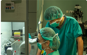 ResearchResearch is one of the cornerstones of Centro Médico Teknon.
 Personalised Follow-up ProgrammeWe accompany you throughout your medical journey. We organise your appointments and tests.
 Patient Quality and SafetyOur management models are based on the most stringent national and international standards.
- Teknon Healthcare Service Areas
- News
- News
 NewsKeep abreast of the events at Centro Médico Teknon. Visit our News section.
 AgendaHere we post upcoming events and discussions on relevant health topics. Visit our Agenda section to see what’s up.
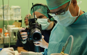 VideosThis section contains an extensive collection of videos related to our specialities.
 PodcastOur specialists discuss current medical topics, innovative treatments, health advice and patient experience.
 Health content
- News
- Blog

- Medical directory
- Specialities
- Specialised Units
- Teknon Cardiology Institute
- Obesity Unit
- Teknon Oncology Institute
- Bloodless Medicine and Surgery Unit
- Teknon Institute of Neurosciences
- Traffic Accident Care Unit
- Teknon Pulmonology Unit
- Maritime Medicine Unit
- Institute of Tissue Regenerative Therapy
- Teknon Pain Treatment Unit
- Teknon Tennis Clinic
- Systemic Inflammatory and Autoimmune Diseases Unit
- Assisted Reproduction Unit
- Sleep Unit
- Unidad de Síndromes de Sensibilización Central
- Specialised Units
- Diagnostics
- Diagnostic tests
- Diagnostic ImagingDiagnostic and interventional scans.
- Anatomical pathology laboratoryAllows to obtain a second opinion from renowned specialists.
- Clinical Analysis LaboratoryComprehensive service in the clinical area.
- EndoscopyAn accurate diagnosis without conventional surgery.
- ElectrophysiologyFunctional exploration of the central nervous system.
- ElectromyographyClinical and neurophysiological evaluation of neuromuscular pathology.
- DensitometryDiagnostic technique for checking bone mineral density.
- UrodynamicsDiagnosis of urination disorders and incontinence.
- Medical check-ups
- GeneralThe most intelligent health check-up
- FullA comprehensive examination of your health
- Full PlusOur most exclusive check-up
- TravellersWhen travelling, your health is also part of your luggage
- SportA thorough review to boost your performance
- CardiologyGood news is knowing your heart is under control
- For companiesA tool that enhances employee satisfaction, productivity, and loyalty
- Diagnostic tests
- Our centre
- Teknon Healthcare Service Areas
- InpatientBright, functional and fully equipped rooms.
- Semi-critical Care UnitEquipped with technology for diagnoses and treatments that require special care.
- Healthy Nutrition ProgrammeWe want to improve people’s health, which is why we promote healthy, conscious and sustainable nutrition at our hospitals.
- NursingOver 400 professionals.
- Emergency DepartmentUninterrupted operation, 24/7 at your service.
- Exclusivity / Teknon ClubCommitted to superior and individualised service, we furnish a full range of services in conjunction with our medical and healthcare support.
- Surgical AreaA total of twenty (20) operating theatres, 12 of which are equipped for high-risk surgery.
- International ProgrammeA programme agent will provide you with comprehensive, personalised support
- Healthcare Ethics CommitteeGuidance for citizens and professionals in cases of moral conflicts.
- ICU-CCUMultipurpose unit incorporating treatment cubicles equipped with modern monitoring systems.
- Patient servicesAvailable to all our patients and their companions.
- ResearchResearch is one of the cornerstones of Centro Médico Teknon.
- Personalised Follow-up ProgrammeWe accompany you throughout your medical journey. We organise your appointments and tests.
- Patient Quality and SafetyOur management models are based on the most stringent national and international standards.
- Teknon Healthcare Service Areas
- News
- News
- NewsKeep abreast of the events at Centro Médico Teknon. Visit our News section.
- AgendaHere we post upcoming events and discussions on relevant health topics. Visit our Agenda section to see what’s up.
- VideosThis section contains an extensive collection of videos related to our specialities.
- PodcastOur specialists discuss current medical topics, innovative treatments, health advice and patient experience.
- Health content
- News
- Blog


- Specialised Units
 Teknon Institute of Neurosciences
- Datos de interés
- Safe brain surgery
Safe brain surgery
The Institute of Neurosciences has developed a series of integrated procedures to ensure maximum safety in its brain surgery procedures.
- Before the procedure. A proper planning for brain surgery enables the integration of an increasing amount of anatomical, physiological, and functional information that can be incorporated into a stereoscopic 3D model of the skull and brain. This model can be viewed in virtual/augmented reality and in 2D, and can also be printed on 3D printers. We can segment a tumour or brain resection, simulate a cranial opening, take rectilinear and curvilinear measurements, integrate physiological information about the tumour or brain (PETs - positron emission tomography activity maps -, SPECT and SISCOM (especially in epilepsy surgery), MR perfusion sequences), functional (functional MRI maps of tasks and resting state, and TMS), as well as morphological connectivity sequences (HARDI tractography).
Ultimately, the goal is to obtain optimal safety margins and surgical approaches for the removal of tumours and other brain lesions or abnormalities. For example, locations of language and sensory-motor areas, as well as their fibres and connectivity tracts around or within the planned resection.
- During the procedure. Advanced neuronavigation allows all of the aforementioned planning elements to be integrated, co-registered in the operating theatre with each patient's actual anatomy, and visualised during surgery. Anatomical elements in the surgical field often shift, which in extreme cases can be up to 2 cm, referred to as cerebral ‘shift’. To correct this, intraoperative ultrasound is used, which can be co-registered with the neuronavigator image. To further minimise the risk to functions such as motor skills or sensitivity, neurophysiological monitoring systems are used with the patient under general anaesthesia. To minimise the impact on language function, the patient undergoes surgery while awake, which allows this function to be mapped in the cerebral cortex and also subcortically during removal in the white matter, while a neuropsychologist with highly specific training monitors this function. Using these techniques, Dr Conesa has operated on more than 700 patients since 1989, with a morbidity rate of less than 4% for these procedures.
In addition to these functional safety features, the Institute of Neurosciences has a complete Kinevo (Zeiss) robotic platform, which also enables an additional Quevo support endoscope with integrated vision directly on the surgical microscope viewer or the built-in screens. In addition, brain tumours can be marked with red fluorescence using 5-ALA (Gliolan®) or yellow fluorescence using fluorescein. The cerebral vascular network can also be viewed using fluorescence. This 4K platform also allows these surgeries to be performed in exoscopic mode and surgical recordings of the highest quality to be made.
The laser brain ablation system (Visualase by Medtronic) has ushered in a paradigm shift in brain surgery. Access to the target lesion is achieved through a minimal cranial opening of 1.6 mm. The precision in positioning the laser probe is optimal with the innovative Leksell Vantage stereotactic system, which is compatible with MRI as it is made of carbon. The functional safety and efficacy of ablation on the target tissue is observed online in the MRI room by superimposing the synchronous recording of highly detailed morphological sequences with high-precision sequences in the recording of temperature changes (thermographic).
Finally, the possibility of performing surgeries in the hybrid operating theatre allows for mixed endovascular and microsurgical procedures to be carried out on cerebral aneurysms, cerebral arteriovenous malformations (AVMs) and arteriovenous fistulas. It also allows verification of the microsurgical closure of the operated aneurysm and the excised AVM or interrupted AVF.