

- Medical directory
- Specialities
- Specialised Units
 Teknon Cardiology Institute
 Obesity Unit
 Teknon Oncology Institute
 Bloodless Medicine and Surgery Unit
 Teknon Institute of Neurosciences
 Traffic Accident Care Unit
 Teknon Pulmonology Unit
 Maritime Medicine Unit
 Institute of Tissue Regenerative Therapy
 Teknon Pain Treatment Unit
 Teknon Tennis Clinic
 Systemic Inflammatory and Autoimmune Diseases Unit
 Assisted Reproduction Unit
 Sleep Unit
 Unidad de Síndromes de Sensibilización Central
- Specialised Units
- Diagnostics
- Diagnostic tests
 Diagnostic ImagingDiagnostic and interventional scans.
 Anatomical pathology laboratoryAllows to obtain a second opinion from renowned specialists.
 Clinical Analysis LaboratoryComprehensive service in the clinical area.
 EndoscopyAn accurate diagnosis without conventional surgery.
 ElectrophysiologyFunctional exploration of the central nervous system.
 ElectromyographyClinical and neurophysiological evaluation of neuromuscular pathology.
 DensitometryDiagnostic technique for checking bone mineral density.
 UrodynamicsDiagnosis of urination disorders and incontinence.
- Medical check-ups

 GeneralThe most intelligent health check-up
 FullA comprehensive examination of your health
 Full PlusOur most exclusive check-up
 TravellersWhen travelling, your health is also part of your luggage
 SportA thorough review to boost your performance
 CardiologyGood news is knowing your heart is under control
 For companiesA tool that enhances employee satisfaction, productivity, and loyalty
- Diagnostic tests
- Our centre
- Teknon Healthcare Service Areas
 InpatientBright, functional and fully equipped rooms.
 Semi-critical Care UnitEquipped with technology for diagnoses and treatments that require special care.
 Healthy Nutrition ProgrammeWe want to improve people’s health, which is why we promote healthy, conscious and sustainable nutrition at our hospitals.
 NursingOver 400 professionals.
 Emergency DepartmentUninterrupted operation, 24/7 at your service.
 Exclusivity / Teknon ClubCommitted to superior and individualised service, we furnish a full range of services in conjunction with our medical and healthcare support.
 Surgical AreaA total of twenty (20) operating theatres, 12 of which are equipped for high-risk surgery.
 International ProgrammeA programme agent will provide you with comprehensive, personalised support
 Healthcare Ethics CommitteeGuidance for citizens and professionals in cases of moral conflicts.
 ICU-CCUMultipurpose unit incorporating treatment cubicles equipped with modern monitoring systems.
 Patient servicesAvailable to all our patients and their companions.
 ResearchResearch is one of the cornerstones of Centro Médico Teknon.
 Personalised Follow-up ProgrammeWe accompany you throughout your medical journey. We organise your appointments and tests.
 Patient Quality and SafetyOur management models are based on the most stringent national and international standards.
- Teknon Healthcare Service Areas
- News
- News
 NewsKeep abreast of the events at Centro Médico Teknon. Visit our News section.
 AgendaHere we post upcoming events and discussions on relevant health topics. Visit our Agenda section to see what’s up.
 VideosThis section contains an extensive collection of videos related to our specialities.
 PodcastOur specialists discuss current medical topics, innovative treatments, health advice and patient experience.
 Health content
- News
- Blog

- Medical directory
- Specialities
- Specialised Units
- Teknon Cardiology Institute
- Obesity Unit
- Teknon Oncology Institute
- Bloodless Medicine and Surgery Unit
- Teknon Institute of Neurosciences
- Traffic Accident Care Unit
- Teknon Pulmonology Unit
- Maritime Medicine Unit
- Institute of Tissue Regenerative Therapy
- Teknon Pain Treatment Unit
- Teknon Tennis Clinic
- Systemic Inflammatory and Autoimmune Diseases Unit
- Assisted Reproduction Unit
- Sleep Unit
- Unidad de Síndromes de Sensibilización Central
- Specialised Units
- Diagnostics
- Diagnostic tests
- Diagnostic ImagingDiagnostic and interventional scans.
- Anatomical pathology laboratoryAllows to obtain a second opinion from renowned specialists.
- Clinical Analysis LaboratoryComprehensive service in the clinical area.
- EndoscopyAn accurate diagnosis without conventional surgery.
- ElectrophysiologyFunctional exploration of the central nervous system.
- ElectromyographyClinical and neurophysiological evaluation of neuromuscular pathology.
- DensitometryDiagnostic technique for checking bone mineral density.
- UrodynamicsDiagnosis of urination disorders and incontinence.
- Medical check-ups
- GeneralThe most intelligent health check-up
- FullA comprehensive examination of your health
- Full PlusOur most exclusive check-up
- TravellersWhen travelling, your health is also part of your luggage
- SportA thorough review to boost your performance
- CardiologyGood news is knowing your heart is under control
- For companiesA tool that enhances employee satisfaction, productivity, and loyalty
- Diagnostic tests
- Our centre
- Teknon Healthcare Service Areas
- InpatientBright, functional and fully equipped rooms.
- Semi-critical Care UnitEquipped with technology for diagnoses and treatments that require special care.
- Healthy Nutrition ProgrammeWe want to improve people’s health, which is why we promote healthy, conscious and sustainable nutrition at our hospitals.
- NursingOver 400 professionals.
- Emergency DepartmentUninterrupted operation, 24/7 at your service.
- Exclusivity / Teknon ClubCommitted to superior and individualised service, we furnish a full range of services in conjunction with our medical and healthcare support.
- Surgical AreaA total of twenty (20) operating theatres, 12 of which are equipped for high-risk surgery.
- International ProgrammeA programme agent will provide you with comprehensive, personalised support
- Healthcare Ethics CommitteeGuidance for citizens and professionals in cases of moral conflicts.
- ICU-CCUMultipurpose unit incorporating treatment cubicles equipped with modern monitoring systems.
- Patient servicesAvailable to all our patients and their companions.
- ResearchResearch is one of the cornerstones of Centro Médico Teknon.
- Personalised Follow-up ProgrammeWe accompany you throughout your medical journey. We organise your appointments and tests.
- Patient Quality and SafetyOur management models are based on the most stringent national and international standards.
- Teknon Healthcare Service Areas
- News
- News
- NewsKeep abreast of the events at Centro Médico Teknon. Visit our News section.
- AgendaHere we post upcoming events and discussions on relevant health topics. Visit our Agenda section to see what’s up.
- VideosThis section contains an extensive collection of videos related to our specialities.
- PodcastOur specialists discuss current medical topics, innovative treatments, health advice and patient experience.
- Health content
- News
- Blog


- Specialities
 Bardají Bofill Manel
- Servicios
- Head and neck
- Cysts
- Cervicofacial cysts
- Mixed Parotid Tumor
- Centro Médico TeknonCentre Mèdic Quirónsalud Badalona

What is it?
The mixed tumor or pleomorphic adenoma is applied to all tumors of the salivary and skin glands containing mesenchymal epithelial elements. 90% of cases are mainly located in the parotid; 7%, in the submaxillary gland and palatine vault; 1% in the sublingual glands and the remaining 2-3% in the accessory, sweat and lacrimal glands, having been described in order of frequency in jugal mucosa, neck, lips, cheeks, eyelids, etc.
The cutaneous localization takes the form of subcutaneous or infrared nodules of firm consistency, well delimited, smooth or lobly surface, not adhered to the superficial or deep and mobile planes and although they can settle anywhere on the body, they do so mainly on the face, head and neck. Subjectively they are asymptomatic and the discomfort they cause is due to their size.
These tumors have their origin in the sweat glands and derive from the epithelial and myoepithelial cells of these organs and by their histopathological appearance resemble salivary glands.
The mixed parotid tumor is a relatively frequent lesion, of solid or rubbery consistency that is located preauricular or subauricular, is slow growing, not painful, and does not produce facial paresis. The lesion can form anywhere in the parotid, usually in the superficial lobe, sometimes affecting the deepest portion of the gland and the tumor can enter through the stylomaxillary tunnel and enter the parapharyngeal space. In the submaxillary gland the mixed tumor presents as a painless swelling and palpation of a firm lump is more frequent in the outer portion of the gland. Mixed tumors are benign although there are two malignant variants, carcinoma ex pleomorphic adenoma (where the epithelial component usually causes undifferentiated carcinoma) and true malignant mixed tumor.
The capsule of mixed benign tumors varies greatly in thickness and continuity. There may be tumor cells on the surface or arranged as outgrowths outside the capsule. This multicentric proliferation imposes a wide surgical excision that includes the normal gland.
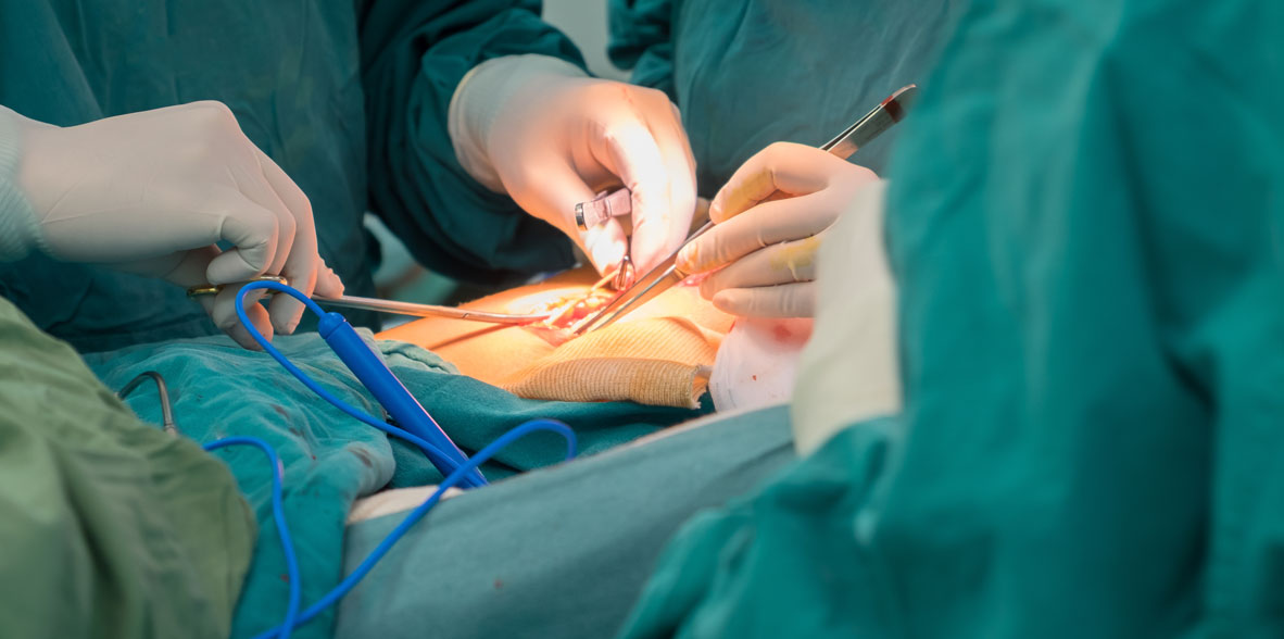
The clinical diagnosis is completed with the aspiration puncture that gives us the diagnosis and the computerized axial tomography that defines its extension and neighborly relations. The treatment is surgical and always requires a parathyroidectomy-type approach, identifying the facial nerve at its exit from the stylomastoid foramen, following it to its terminal branches, if the lesion is limited to the superficial lobe, a suprafacial or superficial lobe parotidectomy is performed, but if the tumor affects the deep lobe, we will require total parotidectomy, always having to respect the facial nerve. The lesion that affects the submaxillary gland must be cleaved in its entirety including the tumor preserving the mandibular branch of the facial. The recurrence of these lesions is usually due to insufficient resection, and must include in the second wide margins of tissue, to prevent additional recurrences, in these patients it may be inevitable to sacrifice branches of the facial nerve, in certain cases suture or nerve graft is indicated.
 Parotid Pleomorphic Adenoma, we clearly observe the normal parotid parenchyma and the mixed tumor (white) that bulges in the subcutaneous
- Congenital parotid cyst
Congenital parotid cysts can appear in infancy or at older ages. They are characterized by dilation of the main canalicular system and formation of multilocular cystic areas. Its extension can be recognized broadly to inspection and palpation, we can indicate sialography, if the ducts are permeable and communicate with the cysts, the contrast medium delineates the cystic spaces, although the computerized axial tomography will reveal the cystic character of the lesion. At first there is clear saliva trapped if there is no infection. Over time there is a risk of chronic infection, with surgical excision with facial preservation being the appropriate treatment.