

menuPrincipal
- Medical directory
- Specialities
- Specialised Units
 Teknon Cardiology Institute
 Obesity Unit
 Teknon Oncology Institute
 Bloodless Medicine and Surgery Unit
 Teknon Institute of Neurosciences
 Traffic Accident Care Unit
 Teknon Pulmonology Unit
 Maritime Medicine Unit
 Institute of Tissue Regenerative Therapy
 Teknon Pain Treatment Unit
 Teknon Tennis Clinic
 Systemic Inflammatory and Autoimmune Diseases Unit
 Assisted Reproduction Unit
 Sleep Unit
 Unidad de Síndromes de Sensibilización Central
- Specialised Units
- Diagnostics
- Diagnostic tests
 Diagnostic ImagingDiagnostic and interventional scans.
 Anatomical pathology laboratoryAllows to obtain a second opinion from renowned specialists.
 Clinical Analysis LaboratoryComprehensive service in the clinical area.
 EndoscopyAn accurate diagnosis without conventional surgery.
 ElectrophysiologyFunctional exploration of the central nervous system.
 ElectromyographyClinical and neurophysiological evaluation of neuromuscular pathology.
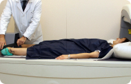 DensitometryDiagnostic technique for checking bone mineral density.
 UrodynamicsDiagnosis of urination disorders and incontinence.
- Medical check-ups

 GeneralThe most intelligent health check-up
 FullA comprehensive examination of your health
 Full PlusOur most exclusive check-up
 TravellersWhen travelling, your health is also part of your luggage
 SportA thorough review to boost your performance
 CardiologyGood news is knowing your heart is under control
 For companiesA tool that enhances employee satisfaction, productivity, and loyalty
- Diagnostic tests
- Our centre
- Teknon Healthcare Service Areas
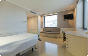 InpatientBright, functional and fully equipped rooms.
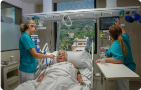 Semi-critical Care UnitEquipped with technology for diagnoses and treatments that require special care.
 Healthy Nutrition ProgrammeWe want to improve people’s health, which is why we promote healthy, conscious and sustainable nutrition at our hospitals.
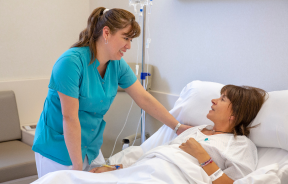 NursingOver 400 professionals.
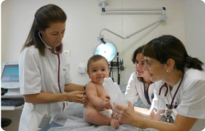 Emergency DepartmentUninterrupted operation, 24/7 at your service.
 Exclusivity / Teknon ClubCommitted to superior and individualised service, we furnish a full range of services in conjunction with our medical and healthcare support.
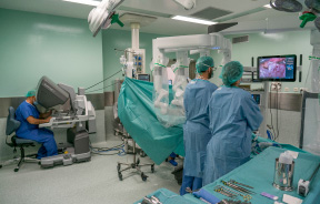 Surgical AreaA total of twenty (20) operating theatres, 12 of which are equipped for high-risk surgery.
 International ProgrammeA programme agent will provide you with comprehensive, personalised support
 Healthcare Ethics CommitteeGuidance for citizens and professionals in cases of moral conflicts.
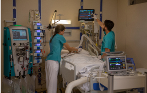 ICU-CCUMultipurpose unit incorporating treatment cubicles equipped with modern monitoring systems.
 Patient servicesAvailable to all our patients and their companions.
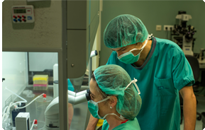 ResearchResearch is one of the cornerstones of Centro Médico Teknon.
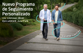 Personalised Follow-up ProgrammeWe accompany you throughout your medical journey. We organise your appointments and tests.
 Patient Quality and SafetyOur management models are based on the most stringent national and international standards.
- Teknon Healthcare Service Areas
- News
- News
 NewsKeep abreast of the events at Centro Médico Teknon. Visit our News section.
 AgendaHere we post upcoming events and discussions on relevant health topics. Visit our Agenda section to see what’s up.
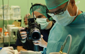 VideosThis section contains an extensive collection of videos related to our specialities.
 PodcastOur specialists discuss current medical topics, innovative treatments, health advice and patient experience.
 Health content
- News
- Blog

menuPrincipal
- Medical directory
- Specialities
- Specialised Units
- Teknon Cardiology Institute
- Obesity Unit
- Teknon Oncology Institute
- Bloodless Medicine and Surgery Unit
- Teknon Institute of Neurosciences
- Traffic Accident Care Unit
- Teknon Pulmonology Unit
- Maritime Medicine Unit
- Institute of Tissue Regenerative Therapy
- Teknon Pain Treatment Unit
- Teknon Tennis Clinic
- Systemic Inflammatory and Autoimmune Diseases Unit
- Assisted Reproduction Unit
- Sleep Unit
- Unidad de Síndromes de Sensibilización Central
- Specialised Units
- Diagnostics
- Diagnostic tests
- Diagnostic ImagingDiagnostic and interventional scans.
- Anatomical pathology laboratoryAllows to obtain a second opinion from renowned specialists.
- Clinical Analysis LaboratoryComprehensive service in the clinical area.
- EndoscopyAn accurate diagnosis without conventional surgery.
- ElectrophysiologyFunctional exploration of the central nervous system.
- ElectromyographyClinical and neurophysiological evaluation of neuromuscular pathology.
- DensitometryDiagnostic technique for checking bone mineral density.
- UrodynamicsDiagnosis of urination disorders and incontinence.
- Medical check-ups
- GeneralThe most intelligent health check-up
- FullA comprehensive examination of your health
- Full PlusOur most exclusive check-up
- TravellersWhen travelling, your health is also part of your luggage
- SportA thorough review to boost your performance
- CardiologyGood news is knowing your heart is under control
- For companiesA tool that enhances employee satisfaction, productivity, and loyalty
- Diagnostic tests
- Our centre
- Teknon Healthcare Service Areas
- InpatientBright, functional and fully equipped rooms.
- Semi-critical Care UnitEquipped with technology for diagnoses and treatments that require special care.
- Healthy Nutrition ProgrammeWe want to improve people’s health, which is why we promote healthy, conscious and sustainable nutrition at our hospitals.
- NursingOver 400 professionals.
- Emergency DepartmentUninterrupted operation, 24/7 at your service.
- Exclusivity / Teknon ClubCommitted to superior and individualised service, we furnish a full range of services in conjunction with our medical and healthcare support.
- Surgical AreaA total of twenty (20) operating theatres, 12 of which are equipped for high-risk surgery.
- International ProgrammeA programme agent will provide you with comprehensive, personalised support
- Healthcare Ethics CommitteeGuidance for citizens and professionals in cases of moral conflicts.
- ICU-CCUMultipurpose unit incorporating treatment cubicles equipped with modern monitoring systems.
- Patient servicesAvailable to all our patients and their companions.
- ResearchResearch is one of the cornerstones of Centro Médico Teknon.
- Personalised Follow-up ProgrammeWe accompany you throughout your medical journey. We organise your appointments and tests.
- Patient Quality and SafetyOur management models are based on the most stringent national and international standards.
- Teknon Healthcare Service Areas
- News
- News
- NewsKeep abreast of the events at Centro Médico Teknon. Visit our News section.
- AgendaHere we post upcoming events and discussions on relevant health topics. Visit our Agenda section to see what’s up.
- VideosThis section contains an extensive collection of videos related to our specialities.
- PodcastOur specialists discuss current medical topics, innovative treatments, health advice and patient experience.
- Health content
- News
- Blog

Language selector

- Specialities
 Bardají Bofill Manel
- Servicios
- Abdomen
- Small intestine
- Jaundice
Consultation area
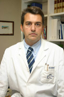
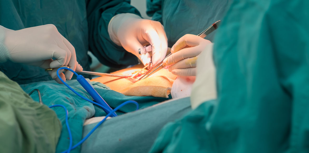
Bardají Bofill ManelGeneral surgery
- Centro Médico TeknonCentre Mèdic Quirónsalud Badalona

Jaundice
Yellowing of the skin, integuments and conjunctiva
- Normal metabolism of bile pigments
Blurrubin is formed from hemoglobin, myoglobin degradation, and by hepatic synthesis. Bilirubin bound to albumin (indirect bilurrubin, insoluble in water (0.3 mg / dl) is transported to the liver, where the albumin is separated and conjugated with glucoronic acid, which is excreted in the intestines, the bacteria reduce bilirubin to urobilinogen (colorless) and urobilin. The excretion of bilirubin in the faeces is 100 to 200 mg/day. Part of the urobilinogen is resorbed in the portal system. - Abnormal metabolism of bile pigments
With normal excretion of bile. Excessive production by hemolysis overloads the liver (increase in indirect bilirubin and urobilinogen); hyperbilirubinemia due to "shunts" and liver defects (Gilbert's disease and Crigler-Najjar syndrome). In all of them there is jaundice, but there is no choluria; that is, the urine is clear. - With disorders in the excretion of bile
Excess conjugated bilirubin is excreted in the urine. Intrahepatic causes. Dubin-Johnson syndrome (normal liver function tests; decreased excretion) drugs (phenothiazines, methyltestosterone); Primary biliary cirrhosis. In hepatitis and cirrhosis (abnormalities in liver function tests) there are deficiencies in conjugation and excretion. Biliary atresia is seen and the stool may be pale, as in mastique (acolia). - Extrahepatic cholestasis
In it there is an anatomical obstacle to the exit of bile from the liver. It is caused by atresia, stricture, stones, tumors, cysts, and parasites. There is an increase in direct and indirect fractions. - Assessment and treatment of jaundice
Jaundice causes clinical manifestations when the bilirubin level is 2.5 mg/100 dl or higher. Yellow dye first appears on the sclera. Anamnesis. The chronological course is important (extrahepatic obstruction has insidious onset); painless jaundice (carcinoma); presence of pain in the right upper quadrant (stones, cholangitis), family history, drug ingestion. Anorexia, fever (hepatitis), contact with infectious patients, history of alcoholism are also observed. - Physical examination
Rashes (drug reactions), skin angiomas (cirrhosis), hepatomegaly and pain to the touch (hepatitis), palpable gallbladder (cancers with extrahepatic obstruction, Courvoisier's law), and ascites. Laboratory studies. They show anemia, increased number of reticulocytes (hemolysis), sickle cells, spherocytes, increased number of leukocytes (cholangitis); stool color (33% of pancreatic carcinomas show positivity to the heme fraction) Direct and indirect bilirubins should be measured. Sometimes there is bilirubinuria, but not with jaundice from unconjugated bilirubins. Alkaline phosphatase (obstructive jaundice) should also be measured, with increased glutamic oxaloacetic and pyruvic transaminases (hepatitis). The increase in prothrombin time and the inadequate response to soluble vitamin K (Aquamenphyton) are data that denote hepatocellular disease. - Radiographic studies
It is advisable to obtain simple x-rays (20% of the stones are opaque). Oral cholecystography is ineffective if bilirubin is 1.8 mg or greater; ultrasonography; endoscopic retrograde cholangiopancreatography (ERCP) (with biopsy), percutaneous transhepatic cholangiography (CPT), computed tomography; Bile duct scan, including 20 mg/100 deciliter bilirubin. - Other studies
Liver biopsy can identify hepatocellular disease, but coagulation must be measured first! Laparotomy is performed in cases of extrahepatic obstruction. Algorithm of obstructive jaundice. If the doctor suspects extrahepatic obstruction, the first study will be ultrasound, which will be followed by the algorithm. - Failure of multiple organ systems
The digestive tract constitutes a barrier against invasions by enteric flora, which can be lost during critical illnesses, especially when there are no intraluminal intestinal nutrients that stimulate mucosal growth. Atrophy causes patency to pathogens, chronic hypermetabolism and finally multiple organ system failure (IMSO). An essential fuel for the enterocyte is intraluminal glutamine.
Patients with IMSO often die from liver or kidney failure, which may be due to humoral mediators triggered by an uncontrolled infection. When a clear source of infection is not proven, it should be attributed to the digestive tract by bacterial translocation. IMSO is best addressed by: (a) detecting and eradicating the infectious source, and (b) providing maximum intensive organ support.
This page contains medical information and guidance for patients, in no way the symptoms or conditions indicated herein, with the respective advice for the prevention of complications and that are related to your person, do not replace the medical evaluation that should be practiced by your family doctor or specialist of your choice. Therefore these concepts should be taken only as medical information or culture. If you require more information, you should go to your trusted family doctor or to the Health Institution closest to your home.