

- Quadre mèdic
- Especialitats
- Unitats especialitzades
 Institut del Cor Teknon
 Unitat d’Obesitat
 Institut Oncològic
 Unitat d’Medicina i cirurgia sense sang
 Institut de Neurociències
 Unitat d’atenció al lesionat de trànsit
 Unitat de Pneumologia
 Unitat de Medicina Marítima
 Institut de Teràpia Regenerativa Tissular
 Unitat de Tractament del Dolor
 Clínica del Tennis
 Unitat de malalties inflamatòries i autoimmunes sistèmiques
 Unitat de Reproducció Assistida
 Unitat del Son
 Unitat de Síndromes de Sensibilització Central
- Unitats especialitzades
- Àrea diagnòstica
- Proves diagnòstiques
 Diagnòstic per la ImatgeExploracions diagnòstiques i intervencionistes.
 Laboratori d’anatomia patològicaDisposa d’una segona opinió per part d’especialistes.
 Laboratori d’Anàlisis ClíniquesServei integral a l’àrea clínica.
 EndoscòpiaDiagnòstic precís sense utilitzar la cirurgia.
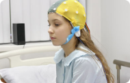 ElectrofisiologiaExploració funcional del Sistema Nerviós Central.
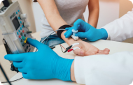 ElectromiografíaAvaluació clínica i neurofisiològica de la patologia neuromuscular.
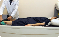 DensitometriaTècnica diagnòstica per comprovar la densitat mineral de l’os.
 UrodinàmiaDiagnòstic dels trastorns de micció i incontinència.
- Revisions mèdiques

 GeneralUn control intel·ligent de la teva salut.
 CompletUn examen exhaustiu de la teva salut.
 Complet PlusLa nostra revisió mèdica més exhaustiva.
 ViatgersSi vols fer un viatge, la teva salut forma part de l’equipatge.
 EsportiuUna revisió a fons per a potenciar el teu rendiment.
 CardiològicUna bona notícia és saber que el teu cor està sota control.
 Per a empresesUna eina que potencia la satisfacció, productivitat i fidelització del treballador.
- Proves diagnòstiques
- El nostre centre
- Entorn assistencial
 HospitalitzacióAmb habitacions lluminoses, funcionals i completament equipades.
 Unitat de SemicriticsDotada de tecnologia per al diagnòstic i tractament que requereixen una vigilància i cura especial.
 Programa d’Alimentació SaludableVolem millorar la salut de les persones, per això promovem una alimentació saludable, conscient i sostenible als nostres hospitals.
 InfermeriaUn equip de més de 400 professionals.
 Servei d’Urgències24 hores al dia al teu servei sense interrupcions.
 Àrea Pacient PrivatBasat en l’alta qualitat i el servei personalitzat, oferim un conjunt de serveis que complementen les cures mèdico-assistencials
 L’Àrea QuirúrgicaDisposa de 20 quiròfans, 12 d’ells preparats per a la cirurgia d’alt risc.
 International programUn assistent d’aquest programa us oferirà atenció integral i personalitzada
 Comitè d’Ètica AssistencialAjuda els ciutadans i als professionals de la salut a orienta la seva actuació en casos de conflictes morals.
 UCI-UCUnitat polivalent que disposa de boxes completament equipats amb el sistema més modern de monitorització.
 Atenció al PacientA disposició de tots els pacients i acompanyants del centre.
 InvestigacióLa investigació constitueix un dels pilars bàsics de Centre Mèdic Teknon.
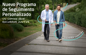 Programa de Seguiment PersonalitzatT’acompanyem durant el teu procés mèdic. Organitzem i agendem les teves cites i proves.
 Qualitat i Seguretat del PacientAdoptem models de gestió basats en els estàndards més exigents nacionals i internacionals.
- Entorn assistencial
- Actualitat
- Actualitat
 NotíciesConeix el que està passant a Centro Médico Teknon. Consulta la nostra secció de notícies.
 AgendaPots trobar tots els esdeveniments que hem organitzat sobre salut i aquells temes d’actualitat que et poden interessar. Accedeix a la nostra agenda d’activitats.
 VídeosEn aquesta secció trobaràs una àmplia col·lecció de vídeos relacionats amb les nostres especialitats.
 PodcastTemes mèdics d’actualitat, tractaments innovadors, consells de salut y experiències de pacients abordabats pels nostres especialistes.
 Continguts de salut
- Actualitat
- Blog

- Quadre mèdic
- Especialitats
- Unitats especialitzades
- Institut del Cor Teknon
- Unitat d’Obesitat
- Institut Oncològic
- Unitat d’Medicina i cirurgia sense sang
- Institut de Neurociències
- Unitat d’atenció al lesionat de trànsit
- Unitat de Pneumologia
- Unitat de Medicina Marítima
- Institut de Teràpia Regenerativa Tissular
- Unitat de Tractament del Dolor
- Clínica del Tennis
- Unitat de malalties inflamatòries i autoimmunes sistèmiques
- Unitat de Reproducció Assistida
- Unitat del Son
- Unitat de Síndromes de Sensibilització Central
- Unitats especialitzades
- Àrea diagnòstica
- Proves diagnòstiques
- Diagnòstic per la ImatgeExploracions diagnòstiques i intervencionistes.
- Laboratori d’anatomia patològicaDisposa d’una segona opinió per part d’especialistes.
- Laboratori d’Anàlisis ClíniquesServei integral a l’àrea clínica.
- EndoscòpiaDiagnòstic precís sense utilitzar la cirurgia.
- ElectrofisiologiaExploració funcional del Sistema Nerviós Central.
- ElectromiografíaAvaluació clínica i neurofisiològica de la patologia neuromuscular.
- DensitometriaTècnica diagnòstica per comprovar la densitat mineral de l’os.
- UrodinàmiaDiagnòstic dels trastorns de micció i incontinència.
- Revisions mèdiques
- GeneralUn control intel·ligent de la teva salut.
- CompletUn examen exhaustiu de la teva salut.
- Complet PlusLa nostra revisió mèdica més exhaustiva.
- ViatgersSi vols fer un viatge, la teva salut forma part de l’equipatge.
- EsportiuUna revisió a fons per a potenciar el teu rendiment.
- CardiològicUna bona notícia és saber que el teu cor està sota control.
- Per a empresesUna eina que potencia la satisfacció, productivitat i fidelització del treballador.
- Proves diagnòstiques
- El nostre centre
- Entorn assistencial
- HospitalitzacióAmb habitacions lluminoses, funcionals i completament equipades.
- Unitat de SemicriticsDotada de tecnologia per al diagnòstic i tractament que requereixen una vigilància i cura especial.
- Programa d’Alimentació SaludableVolem millorar la salut de les persones, per això promovem una alimentació saludable, conscient i sostenible als nostres hospitals.
- InfermeriaUn equip de més de 400 professionals.
- Servei d’Urgències24 hores al dia al teu servei sense interrupcions.
- Àrea Pacient PrivatBasat en l’alta qualitat i el servei personalitzat, oferim un conjunt de serveis que complementen les cures mèdico-assistencials
- L’Àrea QuirúrgicaDisposa de 20 quiròfans, 12 d’ells preparats per a la cirurgia d’alt risc.
- International programUn assistent d’aquest programa us oferirà atenció integral i personalitzada
- Comitè d’Ètica AssistencialAjuda els ciutadans i als professionals de la salut a orienta la seva actuació en casos de conflictes morals.
- UCI-UCUnitat polivalent que disposa de boxes completament equipats amb el sistema més modern de monitorització.
- Atenció al PacientA disposició de tots els pacients i acompanyants del centre.
- InvestigacióLa investigació constitueix un dels pilars bàsics de Centre Mèdic Teknon.
- Programa de Seguiment PersonalitzatT’acompanyem durant el teu procés mèdic. Organitzem i agendem les teves cites i proves.
- Qualitat i Seguretat del PacientAdoptem models de gestió basats en els estàndards més exigents nacionals i internacionals.
- Entorn assistencial
- Actualitat
- Actualitat
- NotíciesConeix el que està passant a Centro Médico Teknon. Consulta la nostra secció de notícies.
- AgendaPots trobar tots els esdeveniments que hem organitzat sobre salut i aquells temes d’actualitat que et poden interessar. Accedeix a la nostra agenda d’activitats.
- VídeosEn aquesta secció trobaràs una àmplia col·lecció de vídeos relacionats amb les nostres especialitats.
- PodcastTemes mèdics d’actualitat, tractaments innovadors, consells de salut y experiències de pacients abordabats pels nostres especialistes.
- Continguts de salut
- Actualitat
- Blog


- Especialitats
 Gilete Vicenç
- Servicios
- Artrodesis occipitocervical

Artrodesis occipitocervical
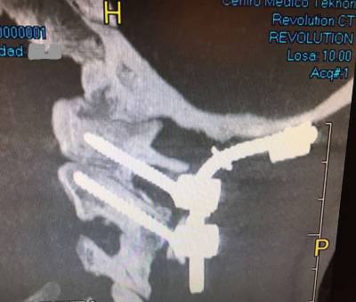 Surgical treatment of craniocervical instability usually consists of the posterior fusion of Occipital, Atlas (C1), and Axis (C2). In the Axis, pedicle screws are usually placed, though depending on the patient’s anatomy, screws can be placed in the isthmus. The Atlas screws are generally placed in the lateral masses. In regard to the fixation of the cranium to above mentioned cervical vertebrae, it can be done mainly in two ways. The most frequent and widespread way is the placement of screws in the Occipital bone. Alternatively, the cranium screws may be placed in the occipital condyle. In any of the two ways, the implanted screws are joined by lateral bars which actually are what gives rigidity to the fusion system. In most cases it is convenient to put bone graft, usually autologous, originated from the iliac crest or from the patient’s own rib. In cases where it is not possible to obtain autologous bone graft, heterologous graft (artificial bone) may also be used.
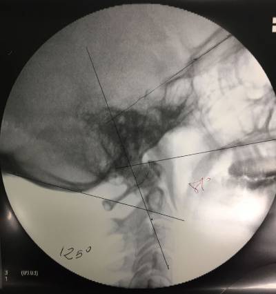
Preoperative and individualized evaluation of each case is extremely important since anomalies in the anatomy and trajectory of the Vertebral Artery should be ruled out. Anatomical differences in regard to dimensions and disposition of vertebral pedicles, lateral masses and other bone elements should also be assessed. The diagnosis can be made by means of an Upright MRI (Magnetic Resonance Imaging) or with a cervical CT scan with 3D reconstruction. Both tests should evaluate the movements of the occipitoatlantoid and atlantoaxial joints. Flexion-extension and cervical rotation on both sides should be evaluated. Lateral cervical x-ray and flexion-extension views can give us complementary information in regards to atlantoaxial instability, although it does not seem indicated as the first choice method of diagnosis. Another diagnostic method used is cervical cineradiology, which records joint(s) movement of the entire occipitocervical, atlantoaxial and subaxial joint system.
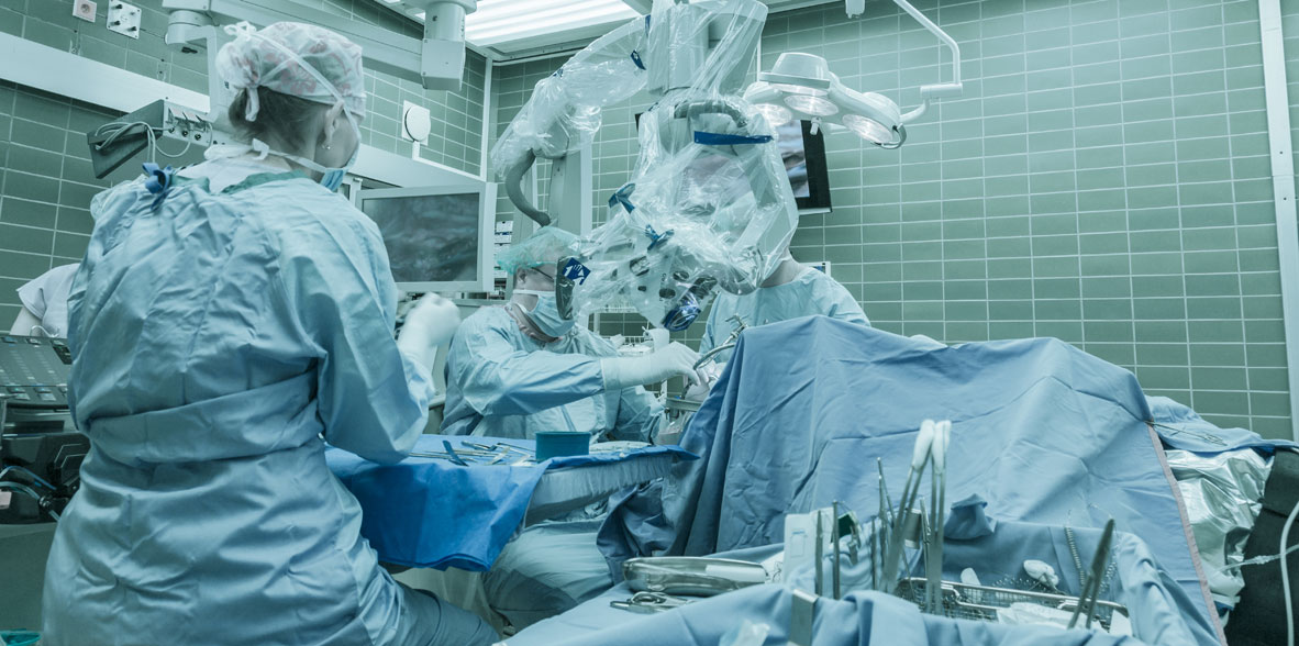
 Once in the Operating Room, surgery is performed under general anesthesia, with Neurophysiological monitoring (SSEP – somatosensory evoked potentials), neuronavigation guidance and intraoperative fluoroscopy guidance.
Thus we control spinal cord and nerves (cranial and cervical) in order to avoid potential damages to these important structures. Neuronavigation assistance guides us all through the surgery, thus it diminishes (though it does not eliminate) the risks while placing the screws for the fusion. Both neurophysiological monitoring and neuronavigation guidance are safety measures for the patient.
Postoperatively, the patient stays at the ICU unit 1 day and then he/she stays in the Neurosurgical Ward. Postoperative hospital stay is usually around 7 days. Wake up and walking begins on the second day after surgery. After hospital discharge, doctors usually control patients at least once a week after discharge on an outpatient basis, to make sure everything is correct before flying back home, thus we recommend to stay in Barcelona after discharge for 10-15 days.
Abbreviations: BDI: basion dens interval, CXA: clivo axial angle, BAI: basion-axial interval, ADI: Atlantoaxial interval
Fuentes:
- Dr. Vicenç Gilete, MD, Neurosurgeon & Spine Surgeon.
- Schulz R, Macchiavello N, Fernández E, Carredano X, Garrido O, Diaz J, Melcher RP. Harms C1-C2 instrumentation technique: anatomo-surgical guide. Spine (Phila Pa 1976). 2011 May 20;36(12):945-50